
Clinical Images: Massive Retinal Melanoma
Authors
Michael T. Lawton, MD†
Robert F. Spetzler, MD†
†Division of Neurological Surgery, Barrow Neurological Institute, St. Joseph’s Hospital and Medical Center, Phoenix, Arizona
Key Words : retinal melanoma
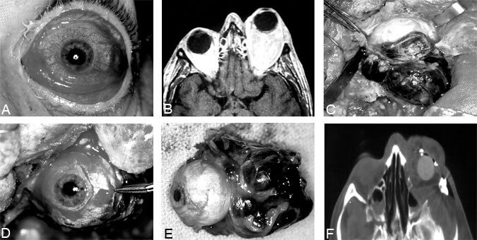
Five years before presenting to this institution, a 41-year-old male developed blurred vision and was diagnosed with an orbital tumor. He decided against treatment and his symptoms progressed to blindness 6 months later. The progression of proptosis and chemosis, however (Panel A), ultimately caused him to seek treatment. Magnetic resonance imaging (Panel B) demonstrated tumor filling the left orbit and compressing the optic nerve. A left frontal craniotomy was performed with orbital exenteration, lateral wall orbitectomy, and orbital reconstruction with a prosthetic globe. The encapsulated tumor was resected en bloc (Panels C-E) and diagnosed as retinal melanoma. Postoperative computed tomography confirmed complete resection of the tumor (Panel F). The patient’s course was uncomplicated, and 4 months after surgery he was fitted with an ocular prosthesis.