Clinical Images: Bullet Extracted from the Craniovertebral Junction via a Transoral Approach
Division of Neurological Surgery, Barrow Neurological Institute, St. Joseph’s Hospital and Medical Center, Phoenix, Arizona
Key Words : bullet, Craniovertebral Junction, pentrating injury, Transoral
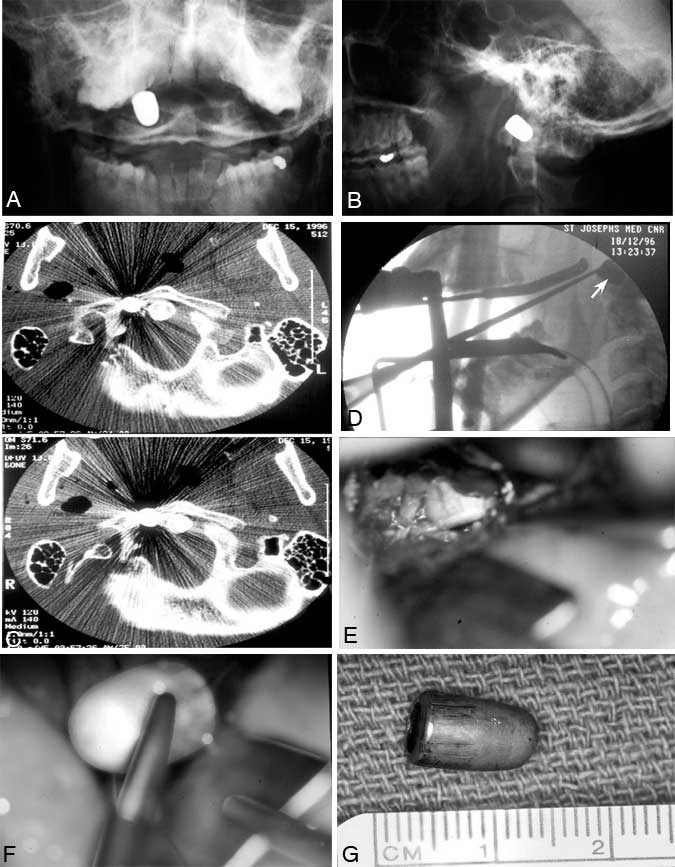
A 28-year-old male sustained a gunshot wound to the face. The bullet entered the right cheek and penetrated the maxillary sinus. Ultimately, the bullet lodged between the anterior ring of C1 and the odontoid process as shown in open-mouth (Panel A) and lateral (Panel B) cervical spine radiographs. The patient was neurologically intact. A computed tomographic scan (axial slice, bone algorithm;Panel C) shows the bullet to the right of the odontoid process.
The patient was placed in a halo brace because of injury to C1 as well as the likelihood of injury to the transverse ligament. He subsequently underwent a transoral approach to remove the bullet. A lateral intraoperative fluoroscopic radiograph of the cervical spine (Panel D) shows the transoral retractor system in place with the tip of a curette used to localize the bullet (arrow). Intraoperative photographs show the bullet slug after the posterior pharynx has been opened (Panel E) and during its removal (Panel F, slightly computer-enhanced). The slug (Panel G) was removed and the patient tolerated the procedure well. Postoperatively, he wore a halo brace for 6 weeks, at which time his dynamic (flexion/extension) cervical spine radiographs were normal (not shown) and the halo brace was removed.