


Approach to Patients with Epilepsy
Joseph Drazkowski, MD
Division of Neurology, Barrow Neurological Institute, St. Joseph’s Hospital and Medical Center, Phoenix, Arizona
Abstract
The etiology and epidemiology of epilepsy are reviewed. The classification of various seizure types is discussed in the context of diagnosing epilepsy. The impact on the life of the person living with epilepsy is also discussed as are patient safety and statutory regulations. An approach to evaluating patients suspected of having epilepsy includes relevant laboratory, electroencephalographic, and diagnostic imaging studies. Finally, special consideration is given to the treatment of the various types of status epilepticus and alcohol withdrawal seizures.
Key Words: epilepsy, seizures
A discussion of epilepsy first requires understanding the terms epilepsy and seizure. A seizure can be defined as a paroxysmal disorder of the central nervous system characterized by abnormal cerebral neuronal discharge that results in an alteration of sensation, motor function, behavior, or consciousness. Epilepsy is the condition of spontaneously recurrent seizures (two or more). Epilepsy syndromes (e.g., juvenile myoclonic epilepsy) combine information on seizure type, electroencephalographic (EEG) pattern, age of onset, clinical signs, and mode of inheritance. A seizure is a symptom that results from a wide variety of known and unknown causes. Epilepsy is a disorder, not a specific disease.
Epidemiology
The incidence of epilepsy in adults is approximately 40 per 100,000, correlating with estimates of more than 100,000 new cases of epilepsy in the United States each year. In developed countries, two-thirds of the cases are idiopathic.13-15 About 1.5 million people in the United States have had a seizure in the last 5 years or are taking an antiepileptic medication.
Consequently, epilepsy is a common disorder. It has been described in documents from as early as 1700 b.c.19 Famous and infamous people have had seizures (Table 1). The incidence rates for seizures in children under 4 years of age and in the elderly are higher than those of the middle years.
Etiology of Seizures
By definition, idiopathic seizures are those with no discernible cause. This definition is a moving target that changes with advances in diagnostic technology. For example, the advent of magnetic resonance (MR) imaging revealed that many previously idiopathic seizures were related to cavernous malformations, dysplasias, or low-grade glial tumors. Despite diagnostic advances, idiopathic remains the most common etiology in any age group. Cryptogenic seizures result from a presumed but not specifically identified lesion. It implies that a lesional cause is present but has not been identified. Together, idiopathic and cryptogenic seizures compose about 66% of new cases of epilepsy.
The remaining seizures are symptomatic with a known cause such as a vascular, congenital, traumatic, neoplastic, degenerative, or infectious etiology. Age also plays a role in etiology of seizure disorders. Among the elderly, etiologies such as stroke and brain tumor are common. Childhood causes include birth trauma, infections, metabolic disorders, and fever. Adult seizures are often caused by head injury, infections, drug and alcohol use, and mesial temporal sclerosis or have a psychogenic origin.
Classification of Seizures
The International Classification of Seizures provides a common language for describing types of seizures. Seizures are classified into three major divisions: (1) partial, (2) generalized, and (3) unclassified (Table 2).
Partial seizures originate in a focal area of the brain. A simple partial seizure does not alter consciousness or recall. Complex partial seizures impair awareness or memory for events that occur during the seizure. Partial seizures may manifest with motor or sensory symptoms. Other more subtle symptoms, such as a sense of déja vu or abnormal thoughts or perceptions or autonomic symptoms such as nausea, flushing, or a sense of heat, are common. Such auras are actually simple partial seizures that progress to complex partial seizures as abnormal electrical activity spreads to wider regions of the brain. In previous versions of the classifications of the epilepsies, complex partial seizures were known as temporal lobe, psychomotor, or limbic seizures.
Complex partial seizures are divided into those with or without an aura or with or without automatic movements. An aura can include diverse symptoms such as nausea, focal motor or sensory symptoms, visual changes, or even emotional lability such as a sense of fear or confusion. Complex partial seizures can secondarily generalize into tonic-clonic seizures. A complex partial seizure without aura or automatisms can be difficult to recognize, manifesting only with an interruption in ongoing activity and awareness.
Generalized seizures have an apparent onset over wide areas of the brain and have no focal onset as recorded by scalp EEG. Typical seizures of generalized onset include absence (petit mal), tonic-clonic (grand mal), atonic (drop attacks), and myoclonic seizures.
Simple partial seizures previously were known as focal motor, focal sensory, or Jacksonian seizures. A Jacksonian seizure “marches” from one body part (usually the hand) to the face, trunk, and leg in an organized manner according to the organization of the motor cortex. The classification of seizures is not yet based upon a firm understanding of epilepsy pathophysiology, but it is useful for medical research and systematic communication among physicians. Proper classification of seizures guides medication therapy, specifies the need to look for underlying structural lesions, and provides clues about which patients might be candidates for curative epilepsy surgery.
Diagnosis of Epilepsy
To diagnose epilepsy, one must start with a good story. In general, intermittent episodes associated with stereotyped alterations in sensory or motor function, behavior, or consciousness are consistent with a diagnosis of epilepsy. A good description of an aura, when present, can help both to localize and classify the seizure. A seizure has a clear onset and finish, although the end of some seizures can be obscured by a postictal confusional state. If witnesses observe and report the event carefully, the practitioner can often obtain a clear conception of the event. Phone calls to observers remain the most cost-effective diagnostic technology for epilepsy.
Most seizures last 1 to 2 minutes, but the broader range is 5 seconds to 5 minutes. Although seizures can progress to status epilepticus, most events lasting longer than five minutes (not counting the postictal confusional state or sleep) are not seizures. The appearance of seizures should be stereotyped.
Seizures rarely are triggered. Events precipitated by shouting, emotional upset, eating, cold weather, constipation, and phases of the moon usually are behaviorally induced nonepileptic events. Reflex (triggered) epilepsy, however, is well described for 1 to 5% of the population with epilepsy. Triggers can include flashing lights and other visual stimuli, loud noises, and even certain psychophysiological states. In our epilepsy unit, we have documented (in different patients) seizures triggered by watching a specific cartoon character and by listening to country music by a specific performer.
Epilepsy can be difficult to diagnose even for experts. Imitators of epilepsy are common and broadly can be divided into physiological and psychological imitators (see Table 1 in Nonepileptic Seizures: A Complex Imitator of Epilepsy in this issue). Physiological imitators of epilepsy include syncope, complicated migraine, transient ischemic attacks (TIAs), transient global amnesia, hypoglycemia, sleep disorders, vertigo, movement disorders, and waxing and waning delirium. Syncope can raise the specter of epilepsy if the associated transient brain ischemia produces myoclonic movements or convulsions (convulsive syncope). This situation occurs when the patient is not allowed to recline, and cerebral blood flow is reduced. A complex migraine can manifest with loss of consciousness, visual changes, confusion, and motor and sensory complaints and signs. TIAs also can mimic seizures with associated sensorimotor symptoms, but ischemic-related events typically last longer than seizures. However, a postictal Todd’s paralysis can last for hours.
Psychological imitators of epilepsy include hyperventilation, panic attacks, conversion disorders, and malingering. Hyperventilation can produce sensory symptoms, parasthesias, and a sense of almost fainting if prolonged. Although the symptoms tend to be continuous, they can wax and wane thus imitating recurrent seizure. Panic attacks are transitory and can have a wide variety of motor, sensory, and cognitive symptoms that can mimic seizure. Nonepileptic seizures (pseudoseizures), a form of conversion disorder, are difficult to distinguish from epileptic seizures by simple observation. Adults with nonepileptic seizures often have a history of some form of abuse—physical, emotional, or sexual. Mixed epileptic and nonepileptic seizures present a particularly difficult diagnostic dilemma and usually require video-EEG monitoring to clarify.
Malingering differs from pseudoseizures by the presence of a conscious attempt to deceive the examiner. An epilepsy monitoring unit can often help distinguish true epilepsy from imitators. Simultaneous video and EEG recording of typical events can help provide the diagnosis when a clinical distinction is difficult to make. Video recording of events can be used to educate relatives and significant others about the nature of the event and can help guide appropriate care.
Impact of Epilepsy
Epilepsy imposes a unique set of problems. The threat of injury is a troublesome concern for both patients and practitioners. Those who treat people with epilepsy have all seen serious injury or death caused by seizures. In our clinic alone, patients have been involved in motor vehicle accidents or struck by a train, sustained serious closed head injuries and sidewalk burns that required extensive skin grafting, and drowned or died suddenly without explanation.
Statutory regulations for driving often limit a person with epilepsy from participating in many activities. Depending upon the state, driving can be restricted for variable lengths of time from as little as 3 months to a year. People with epilepsy often cannot hold jobs that involve dangerous machinery, unrestricted working in high places, and commercial driving. Social or personal activity can also be restricted. Swimming or bathing alone can be particularly hazardous. Obtaining a private pilot’s license is almost impossible. Participation in team and individual sports is encouraged by most epileptologists, but children with seizures often are left on the bench or in the bleachers.
Sudden unexplained death is a serious problem associated with epilepsy but fortunately limited to a small percentage of patients. Sudden unexplained death may be caused by cardiac arrhythmia or respiratory insufficiency, but the pathophysiology is unknown and this tragedy cannot be predicted reliably.
Medication can also affect individuals with epilepsy adversely. Common side effects of medicines include memory loss, fatigue, lack of coordination, tremor, weight gain, sexual dysfunction, and even long-term physical effects such as cerebellar atrophy and hepatic dysfunction. The cost of many new medications can be staggering, often leading to financial hardship for patient and family. Costs are particularly difficult for unemployed individuals with epilepsy.
Evaluation of the Person with Epilepsy
A patient with suspected seizures should receive a standard medical evaluation, including history, physical examination, and laboratory testing, to rule out important possible etiologies for the seizures such as hypoxia, hypoglycemia, hypocalcemia, hyponatremia, hypomagnesemia, uremia, liver failure, medication intoxication, and alcohol or recreational drug abuse. A thorough history is key to the evaluation of a person with suspected epilepsy. The single most useful step in the diagnosis of epilepsy is to acquire a detailed description of the event believed to be a seizure.21 Although each case is unique, many factors can help determine if a diagnosis of epilepsy is appropriate (Table 3).
People with epilepsy often have normal physical examinations. Focal findings on their neurological examination, however, may help determine the type of seizure and where it is localized. Body asymmetry, such as differences in the size of the hands or digit, may point to the presence of a developmental injury. Skin lesions such as cafe au lait spots can be associated with inherited phakomatoses. Visceral enlargement can be a clue to certain seizure syndromes or to side effects from medications. Ataxia and nystagmus can be the result of medication.
The interictal EEG plays an important role in the patient suspected of having epilepsy. An EEG showing epileptiform activity sometimes can help define particular seizure syndromes or localize its focus. For example, a generalized spike and wave at 3 Hz is consistent with absence epilepsy and a generalized spike-wave at 5 to 6 Hz with juvenile myoclonic epilepsy. In contrast, focal slowing or epileptiform activity (spikes or sharp waves) suggests partial seizure disorders. The interictal EEG can be normal in patients with epilepsy. Between 12 and 50% of patients who eventually are found to have epilepsy have abnormal EEGs.26 Activation procedures, such as hyperventilation, photic stimulation, sleep, and sleep deprivation, improve the diagnostic yield of standard EEGs. Recording during sleep increases the chance of finding an epileptiform discharge in an additional 40% of those patients whose waking EEGs are normal.6,7,11,12
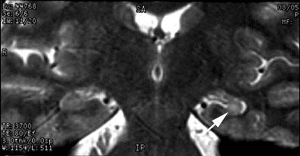
Brain imaging is also essential in the evaluation of individuals with epilepsy. MR imaging is both specific and sensitive for defining structural abnormalities in patients with partial seizure disorders,1,3,4 including neoplasms, vascular malformations, and mesial temporal sclerosis (Fig. 1). Partial epilepsies commonly involve one or both mesial temporal lobes.28 Consequently, high-resolution imaging of the hippocampus and amygdala is very useful for localizing chronic seizure foci of the temporal lobe.18 Arteriovenous malformations, cavernous malformations, focal dysplasias (e.g., heterotopias, migration deficits) and low-grade glial tumors are other common etiologies for seizures and are better detected by MR imaging than computed tomography (CT).2,9
Status Epilepticus
Status epilepticus is defined as a continuous state of seizure or series of seizures with no interval of awakening that lasts longer than 30 minutes. About 150,000 cases of status epilepticus occur each year.8 Convulsive status epilepticus is a medical emergency. The morbidity and mortality rates associated with all types of status epilepticus are high. Each year between 10,000-30,000 people die from status epilepticus or its underlying cause. Neuronal damage caused by status epilepticus is at least partially explained by the excitatory theory of neuronal damage, with a cycle of elevated concentrations of excitatory amino acids and the consequent excessive entry of calcium into the neuron.
Every type of seizure in the International Classification of the Epilepsies has a corresponding type of status epilepticus, which represents prolonged activity of that seizure type. Several classification schemes for status epilepticus have been proposed. One of the simplest schemes consists of (1) partial status epilepticus, (2) generalized convulsive status epilepticus, and (3) nonconvulsive status epilepticus.
Partial Status Epilepticus
Partial status epilepticus represents a prolonged state of focal seizures. In common practice, this term is used to represent prolonged motor or sensory seizures because prolonged complex partial (temporal lobe) status epilepticus is classified under nonconvulsive status epilepticus. The old term for simple partial motor status is epilepsia partialis continua.
Generalized Convulsive Status Epilepticus
Typically, generalized convulsive status epilepticus presents as recurrent convulsions that persist more than 30 minutes without recovery of consciousness between seizures. If allowed to persist, generalized convulsive status epilepticus can progress to a more subtle form associated with only minor motor manifestations. Subtle status epilepticus has a very poor outcome, probably because it is associated with severe injury to the brain.
In a recent Veteran’s Administration (VA) Cooperative Study, about half of the patients responded to therapy for generalized convulsive status epilepticus whereas only 15% responded to therapy if first diagnosed with subtle generalized convulsive status epilepticus.25 Therapeutic outcome was defined as remission of clinical and behavioral EEG responses within 20 minutes. Survivability and morbidity tend to be determined by the underlying cause but can become worse if therapy is inadequate. The mortality rate for generalized convulsive status epilepticus within 30 days is 26%, and it is 65% for subtle status epilepticus.25
EEG plays an important role in diagnosing and determining the response to treatment to generalized convulsive status epilepticus. Overt generalized convulsive status epilepticus is usually clinically obvious, but its subtle form seldom is. Approximately 20% of patients in the VA Cooperative Study of status epilepticus continued to show evidence of status epilepticus on their EEG after their behavioral manifestations stopped. In patients who do not become awake and alert, the therapeutic response should be confirmed by checking an EEG after the overt signs of the seizures have stopped. EEG patterns can show periodic lateralized epileptiform discharges, rhythmic sharp waves or evolving rhythms, spike and wave, continuous spiking, or a burst suppression pattern.
The diverse etiologies of generalized convulsive status epilepticus include trauma; tumor; vascular events; infections; degenerative disease; and toxic, metabolic, and unknown causes. In a review23 of causes of generalized convulsive status epilepticus, trauma was the most common known etiology. When combined, idiopathic and unknown etiologies composed about a third of the cases reviewed.
Nonconvulsive Status Epilepticus
Nonconvulsive status epilepticus can be subdivided into complex partial status epilepticus and absence status epilepticus. The epidemiology of nonconvulsive status epilepticus is still poorly understood, but some studies indicate that 20 to 25% of all cases of status epilepticus are nonconvulsive status epilepticus.5,10 Between 33 and 50% of patients with nonconvulsive status epilepticus present de novo with no history of seizures.22 Common causes of nonconvulsive status epilepticus include vascular events, encephalitis, tumors, congenital malformations, and prior head injury. Status epilepticus can be precipitated in individuals with preexisting epilepsy when their medication is changed or if they fail to comply with their medication regimen.22
During nonconvulsive status epilepticus, the patient’s EEG usually shows either continuous epileptiform activity or recurrent ictal electrographic events in the form of spike-waves, spikes, or evolving rhythms. As the episode continues for hours, increased slowing predominates over spiking. The seizures of patients with cyclical recurrent EEG patterns are more likely to be of temporal origin while those with continuous patterns may have frontal lobe origins.24
Absence status22 is a type of nonconvulsive status epilepticus. The terminology is confusing because a history of absence (petit mal) epilepsy is lacking. Absence status is associated with generalized epileptiform abnormalities on EEG, usually in the form of spike-waves. MR imaging can reveal focal signal abnormalities that can point to focal nonconvulsive status epilepticus.17,29
The clinician needs a high index of suspicion to diagnose nonconvulsive status epilepticus. Unexplained confusion or obtundation could be caused by nonconvulsive status epilepticus but mistaken for psychiatric illness.16,20 Nonconvulsive status epilepticus should be considered in a patient of any age with prolonged unexplained alterations in consciousness, with or without a history of seizure. Whenever a neurological diagnosis of metabolic encephalopathy is considered, the question should arise: Could it be nonconvulsive status epilepticus?
Treatment of Status Epilepticus
The patient with status epilepticus must first be stabilized using standardized medical therapy. The patient’s airway (or intubation if necessary to support respiration or to protect the patient from secretions), blood pressure, and nasal oxygen should be maintained. Electrocardiography should be performed and good intravenous access should be established. A treatable cause of status, such as hypoglycemia, hyponatremia, hypocalcemia, or a toxic overdose, must be identified quickly and treated.
The initial medication used to treat status epilepticus is usually a benzodiazepine—either diazepam (0.25 mg/kg) or lorazepam (0.1 mg/kg). Lorazepam has a more prolonged anticonvulsant effect than diazepam, and its efficacy is also greater.25 After a benzodiazepine has been infused, phenytoin or its water-soluble form, fosphenytoin, is infused to a loading dose of 18 to 20 mg/kg. Phenytoin should be administered no faster than 50 mg/min and fosphenytoin no faster than 150 mg/min. The latter is more expensive but less irritating to tissue, and it may provoke less hypotension and cardiac arrhythmia. If the combination of a benzodiazepine and phenytoin is ineffective, phenobarbital (20 mg/kg) can be given intravenously at a rate no faster than 100 mg/min. If not already in place, intubation is usually necessary at this point. In some patients, pentobarbital, with an intravenous loading dose of 5 mg/kg followed by a maintenance dose titrated to maintain a burst suppression pattern on continuous EEG monitoring, can be more effective than phenobarbital. Midazolam, lidocaine, and propofol are other useful agents when first-line therapies for status epilepticus have failed.
Nonconvulsive status epilepticus should be treated vigorously and expeditiously because postictal cognitive deficits can be profound if it is prolonged. Absence status may respond to benzodiazepines or ethosuximide. Valproic acid can now be used intravenously, but its use has not been established for status epilepticus.
Alcohol Withdrawal Seizures
In a classic article, Victor27 described the alcohol withdrawal syndrome. Mild alcohol withdrawal, with tremors and hallucinations, usually responds to benzodiazepines. Convulsions associated with alcohol withdrawal (rum fits) most often occur during the first 48 hours of abstinence, but they can emerge 12 to 24 hours after a binge. Alcohol withdrawal can also precipitate status epilepticus. This condition is a medical emergency and should be treated the same as generalized convulsive status epilepticus. The occurrence of status epilepticus should prompt further investigation for underlying infection, subdural hematoma, cranial trauma, and a history of epilepsy or other toxic exposure (e.g., cocaine, amphetamines). Withdrawal seizures are usually treated with a benzodiazepine. Oral or rectal paraldehyde has been advocated but is no longer available. Antiepileptic drugs are unnecessary for seizures precipitated only by alcohol; abstinence from alcohol is the therapy of choice. Alcohol abusers also can have chronic epilepsy, in which case compliance with medical therapy becomes a major management dilemma. A careful history about the timing of seizures related to alcohol use may help in the decision process. In each case, therapy must be individualized and depends on the patient’s unique circumstances.
Conclusion
Epilepsy is a relatively common illness and affects people from all walks of life. Seizures have been classified according to the international classification of seizures. The system is useful for describing, medical reporting, and helping to guide therapy. Although seizures have multiple etiologies that are somewhat dependent on a person’s age, large numbers of seizures can be attributed to no definitive known cause. Diagnosing seizures is often challenging, but understanding the various possible imitators is essential for proper therapy. Status epilepticus of all types is a serious medical condition that must be diagnosed quickly and treated effectively. If a diagnosis remains elusive after patients have undergone a complete evaluation, an epilepsy monitoring unit can help establish the diagnosis of epilepsy. People with epilepsy face safety, social, and statutory restrictions in many aspects of their personal and professional lives. Proper diagnosis, understanding the impact of their condition, and effective appropriate treatment should help individuals with epilepsy to maximize the quality of their life.
References
- Berkovic SF, Andermann F, Olivier A, et al: Hippocampal sclerosis in temporal lobe epilepsy demonstrated by magnetic resonance imaging. Ann Neurol 29:175-182, 1991
- Borkovitch A, Chuang S, Norman D: MRI of neuronal migration anomalies. AJNR 8:1009-1017, 1987
- Cascino GD: Commentary: How has neuroimaging improved patient care? Epilepsia 35:103-107, 1994
- Cascino GD, Hirschorn KA, Jack CR, et al: Gadolinium-DTPA-enhanced magnetic resonance imaging in intractable partial epilepsy. Neurology 39:1115-1118, 1989
- Celesia GG: Modern concepts of status epilepticus. JAMA 235:1571-1574, 1976
- Daly DD: Epilepsy and syncope, in Daly DD, Pedley TA (eds): Current Practice of Clinical EEG. New York: Raven, 1990, pp 269-334
- Degen R, Degen HE: Sleep and sleep deprivation in epileptology. Epilepsy Res Suppl 2:235-260, 1991
- DeLorenzo RJ, Pellock JM, Towne AR, et al: Epidemiology of status epilepticus. J Clin Neurophysiol 12:316-325, 1995
- Dodick DW, Cascino GD, Meyer FB: Vascular malformations and intractable epilepsy: Outcome after surgical treatment. Mayo Clin Proc 69:741-745, 1994
- Dunne JW, Summers QA, Stewart-Wynne EG: Non-convulsive status epilepticus: A prospective study in an adult general hospital. Q J Med 62:117-126, 1987
- Ellingson RJ, Wilken K, Bennett DR: Efficacy of sleep deprivation as an activation procedure in epilepsy patients. J Clin Neurophysiol 1:83-101, 1984
- Gibbs EL, Gibbs FA: Diagnostic and localization value of EEG in studies in sleep. Res Publ Assoc Res Nerv Ment Dis 26:366-376, 1947
- Granieri E, Rosati G, Tola R, et al: A descriptive study of epilepsy in the district of Copparo, Italy, 1964-1978. Epilepsia 24:502-514, 1983
- Hauser WA, Kurland LT: The epidemiology of epilepsy in Rochester, Minnesota, 1935 through 1967. Epilepsia 16:1-66, 1975
- Joensen P: Prevalence, incidence, and classification of epilepsy in the Faroes. Acta Neurol Scand 74:150-155, 1986
- Knight RT, Cooper J: Status epilepticus manifesting as reversible Wernicke’s aphasia. Epilepsia 27:301-304, 1986
- Kramer RE, Luders H, Lesser RP, et al: Transient focal abnormalities of neuroimaging studies during focal status epilepticus. Epilepsia 28:528-532, 1987
- Lencz T, McCarthy G, Bronen RA, et al: Quantitative magnetic resonance imaging in temporal lobe epilepsy: Relationship to neuropathology and neuropsychological function. Ann Neurol 31:629-637, 1992
- Scott DF: The History of Epilepsy Therapy: Account of How Medication was Discovered. Pearl River, NY: Parthenon Publishing Group, 1993
- Shorvon SD: Status Epilepticus: Its Clinical Features and Treatment in Children and Adults. Cambridge: Cambridge University Press, 1994
- Stephenson JBP: Fits and Faints. London: MacKeith Press, 1990
- Tomson T, Lindbom U, Nilsson BY: Nonconvulsive status epilepticus in adults: Thirty-two consecutive patients from a general hospital population. Epilepsia 33:829-835, 1992
- Treiman DM: Generalized convulsive status epilepticus in the adult. Epilepsia 34:2-11, 1993
- Treiman DM: Electroclinical features of status epilepticus. J Clin Neurophysiol 12:343-362, 1995
- Treiman DM, Meyers PD, Walton NY, et al: A comparison of four treatments for generalized convulsive status epilepticus. Veterans Affairs Status Epilepticus Cooperative Study Group. N Engl J Med 339:792-798, 1998
- van Donselaar CA, Schimscheimer RJ, Geerts AT, et al: Value of the electroencephalogram in adult patients with untreated idiopathic first seizures. Arch Neurol 49:231-237, 1992
- Victor M: Treatment of alcoholic intoxication and the withdrawal syndrome. A critical analysis of the use of drugs and other forms of therapy. Psychosom Med 28:636-650, 1966
- Williamson PD, Engel J, Jr: Complex partial seizures, in Engel JE, Jr., Pedley TA (eds): Epilepsy. A Comprehensive Textbook. Philadelphia, New York: Lippincott-Raven, 1998, pp 558-559
- Yaffe K, Ferriero D, Barkovich AJ, et al: Reversible MRI abnormalities following seizures. Neurology 45:104-108, 1995