


Burr Holes for Moyamoya Disease
Mark S. Gerber, MD
Robert F. Spetzler, MD
Division of Neurological Surgery, Barrow Neurological Institute, St. Joseph’s Hospital and Medical Center, Phoenix, Arizona
Abstract
Several strategies exist for the treatment of moyamoya disease. A young girl with moyamoya disease was initially treated with a superficial temporal artery onlay procedure and experienced recurrent left-sided ischemic symptoms 2 years later. A perfusion study demonstrated persistent hypoperfusion to the right hemisphere, and a decision was made to place multiple burr holes. The patient’s ischemic symptoms were relieved, and impressive neurovascularization through the burr holes was evident on follow-up angiography.
Key Words: moyamoya disease, burr holes
Moyamoya is an occlusive cerebrovascular disease of unknown etiology. It was first introduced by Suzuki and colleagues in 19667 and named for its angiographic findings of microvascular proliferations that appeared as a puff of smoke. Most of the initial reports of the disease were from Japan; however, cases have since been reported throughout the world. Several surgical options are available for the treatment of moyamoya disease. We report a girl who exhibited impressive revascularization through multiple burr holes after having been previously treated with a superficial temporal artery-to-middle cerebral artery (STA-MCA) onlay procedure.
Illustrative Case
A 12-year-old, left-handed girl initially presented with bitemporal headache, vague left upper extremity weakness and numbness, and intermittent speech difficulties. Cerebral angiography demonstrated bilateral carotid artery occlusion distal to the ophthalmic arteries consistent with moyamoya disease.
In May 1996, the patient underwent a left STA encephaloduroarteriosynangiosis followed by the same procedure on the right side 1 week later. Three-month follow-up angiography showed early evidence of revascularization of both hemispheres. Cerebral angiography 14 months after surgery continued to show subtle improvement compared to the previous study.
The patient was maintained on daily aspirin and remained clinically stable for about 2 years when transient ischemic symptoms involving her left face, shoulder, and arm began to occur with increasing frequency. Magnetic resonance imaging showed no stroke or hemorrhage. A xenon cerebral perfusion study showed poor reserve, especially over the right hemisphere.
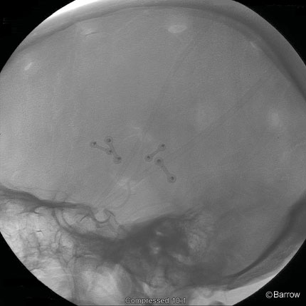
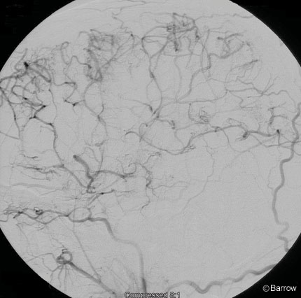
The patient, now 16 years old, was taken to the operating room, and six burr holes were made over the right hemisphere (Fig. 1) as described by Endo et al.[1] Cerebral angiography obtained a year later demonstrated marked leptomeningeal neovascularity at each burr hole (Fig. 2). Because poor perfusion of the left hemisphere persisted, burr holes were placed over the patient’s left hemisphere at this time as well.
Discussion
Although the etiology of moyamoya disease is unknown, it has been associated with Down’s syndrome and von Recklinghausen’s disease. An infectious etiology linked to an abnormal immunological response has also been implicated as a cause of the disease.
Moyamoya tends to manifest during adolescence and again in adults between 30 and 40 years of age. The pediatric population usually becomes symptomatic with transient ischemic attacks, stroke, headache, and seizures. In contrast, adults with moyamoya disease more often present with hemorrhagic events, including subarachnoid hemorrhage.
Classic angiographic findings include occlusion of the supraclinoid internal carotid arteries and absent anterior and MCAs. As a consequence of this progressive occlusion, an impressive network of leptomeningeal and enlarged thalamostriate arteries are seen on angiography.
Medical treatments for moyamoya have included aspirin, steroids, vitamin C, and vasodilators. Numerous surgical procedures have also been described for revascularization of the ischemic brain tissue.
Yasargil[5] performed the first STA-MCA bypass procedure for moyamoya disease in June 1972. This method of revascularization has been used successfully by other surgeons. However, the small size of the vessels limits the utility of the technique in younger children.
In 1973 Goldsmith et al.[2] described a novel surgical approach for the treatment of moyamoya disease. As a result of its known quality of rapid angiogenesis, omentum was selected to be harvested as a free flap, with its vascular supply intact. It was then anastomosed to the STA and laid against the surface of the brain. Several cases were performed with a microsurgical patency rate of approximately 70%.
Henschen[3] first described encephalomyosynangiosis, a procedure in which a flap of the temporalis muscle is freed and laid directly on the surface of the brain. It was thought that over time the muscle would provide small microvascular connections to the pia and brain parenchyma. Postoperative angiograms in children who have undergone this procedure do show improved blood flow to the cerebral cortex. Karasawa and coworkers reported similar angiographic findings after craniotomy and STA-MCA bypass without encephalomyosynangiosis, particularly when meningeal arteries were left intact during surgery.[4]
In 1984 Matsushima and Inaba[6] described encephaloduroarteriosynangiosis. This procedure is somewhat of a combination of an STA-MCA bypass and encephalomyosynangiosis as described above. In encephaloduroarteriosynangiosis, the uninterrupted STA is dissected free and laid over the cortex. This method shortens the operative time and eliminates the need to clip vessels temporarily. The bone flap can be carefully replaced so that blood flow through the STA is not interrupted. Follow-up angiography has confirmed improved blood flow after this procedure.
In 1989 Endo et al.[1] noted that a child with moyamoya who had a frontal burr hole for evacuation of an intracranial hemorrhage had marked neovascularization through the burr hole on follow-up imaging. As a result, they placed frontal burr holes in five children with moyamoya and obtained excellent results in all.
More recently, Suzuki et al.[8] described their experience using a combination of surgical approaches for the treatment of moyamoya disease. Placement of frontal burr holes, in addition to an encephaloduroarteriosynangiosis, encephalomyosynangiosis, or STA-MCA bypass, has provided effective revascularization of ischemic anterior cerebral artery and MCA territories in their pediatric population. These findings, along with those in this case report, highlight the utility of burr holes for the treatment of moyamoya disease.
References
- Endo M, Kawano N, Miyaska Y, et al: Cranial burr hole for revascularization in moyamoya disease. J Neurosurg 71:180-185, 1989
- Goldsmith HS, Chen WF, Duckett SW: Brain vascularization by intact omentum. Arch Surg 106:695-698, 1973
- Henschen C: Revaskularisation des zirkulatorisch geschadigten Gehirns durch Auflage gestielter Muskellappen (Encephalo-myo-synangiose). Langenbecks Arch Chir 264:392, 1950
- Karasawa J, Kikuchi H, Furuse S, et al: Treatment of moyamoya disease with STA-MCA anastomosis. J Neurosurg 49:679-688, 1978
- Krayenbühl HA: The moyamoya syndrome and the neurosurgeon. Surg Neurol 4:353-360, 1975
- Matsushima Y, Inaba Y: Moyamoya disease in children and its surgical treatment. Introduction of a new surgical procedure and its follow-up angiograms. Childs Brain 11:155-170, 1984
- Suzuki J, Takaku A, Asahi M: Evaluation of a group of disorders showing an abnormal vascular network at the brain with a high incidence among the Japanese. II. Follow-up studies by cerebral angiography (transl). No To Shinkei 18:897-908, 1966
- Suzuki Y, Negoro M, Shibuya M, et al: Surgical treatment for pediatric moyamoya disease: Use of the superficial temporal artery for both areas supplied by the anterior and middle cerebral arteries. Neurosurgery 40:324-330, 1997