


Double Sellar Floor: Radiographic Sign for a Pituitary Adenoma
Iman Feiz-Erfan, MD
Giuseppe Lanzino, MD*
William L. White, MD
Division of Neurological Surgery, Barrow Neurological Institute, St. Joseph’s Hospital and Medical Center, Phoenix, Arizona
*Current Address: Department of Neurosurgery; University of Illinois College of Medicine at Peoria; Peoria, Illinois
 Before the introduction of computed tomography and magnetic resonance (MR) imaging, intracranial imaging relied mostly on skull radiographs, pneumencephalography, and cerebral angiography. To diagnose a pituitary adenoma, several signs had to be present on skull radiographs. The size of the sella often could indicate an intrasellar mass. Frequently, pituitary adenomas tend to grow asymmetrically. The sellar floor is then uneven, reflecting uneven enlargement of the bony sella.
On a lateral skull radiograph where the bony lines of the anterior fossa and the sphenoid wings are aligned, a double contoured sellar floor may be appreciated as a radiographic sign for an intrasellar mass lesion causing uneven expansion of the bony sellar floor. However, a double sellar floor can also be caused by parasellar lesions, including aneurysms and meningiomas. Sometimes a double contour of the sellar floor is visible as a variation of normal anatomy and is likely caused by uneven bony growth, a feature called a pseudodouble sellar floor. The double sellar floor along with other bony changes visible on skull radiography, including elevation of the anterior clinoid process and thinning and backward displacement of the dorsum sellae, were once the criteria used to diagnose intrasellar mass lesions on skull radiography.
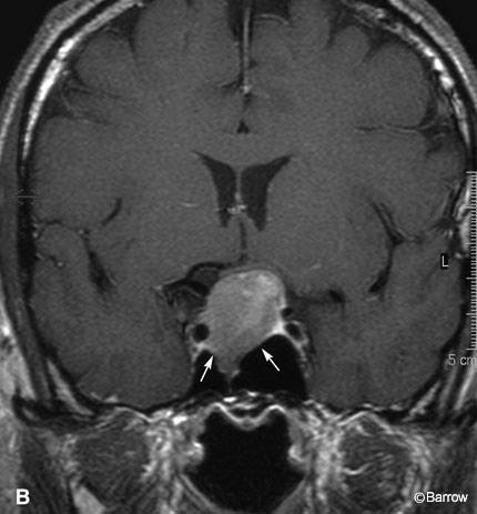
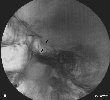 In Panel A an intraoperative aligned lateral skull radiograph of a sella shows a double sellar floor (black arrows) associated with a nonfunctional pituitary macroadenoma in a 57-year-old male who developed symptoms and signs of progressive visual field deficit. The macroadenoma compressing the optic apparatus is also seen on a coronal contrast-enhanced MR image (Panel B). Asymmetric expansion (white arrows) of the sellar floor secondary to uneven tumor growth and sellar compression is evident on the coronal image.