


Brain Aneurysm
Brain Aneurysm Overview
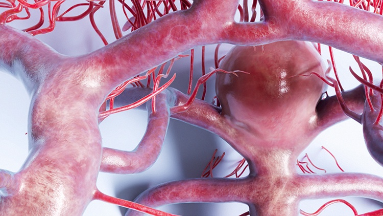
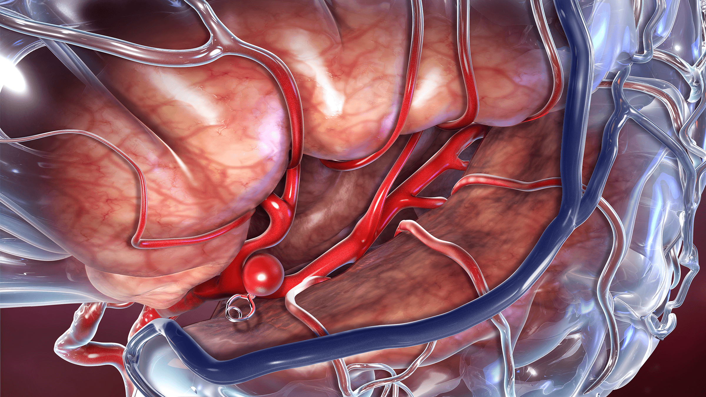
A brain aneurysm, also called a cerebral aneurysm, is a weak spot along a blood vessel in the brain that bulges outward. Often described as balloon-like in appearance, they may burst and bleed into the brain (hemorrhage, or hemorrhagic stroke). This can potentially cause life-threatening complications.
Even if a brain aneurysm does not burst, it can cause other problems by putting pressure on nearby nerves or tissue. Most cerebral aneurysms occur along a ring of interconnected arteries at the base of the brain known as the circle of Willis.
Although some risk factors can increase your risk, the precise cause of brain aneurysm formation is not always known. There is a genetic component, but the specific gene(s) that are involved are still under investigation.
The unique anatomy of an individual can also come into play, that is the way that the arteries in your brain bend and turn. Excessive curves in the arteries can cause hemodynamic stress on the arterial wall, which can degrade the tissue and cause it to weaken, bulge, and bleed.
Anatomy of the Cerebrovascular System
The cerebrovascular system is the network of blood vessels that supply your brain with oxygen and nutrients. The system includes the internal carotid arteries, vertebral arteries, and their branches, which bring blood into the brain, and the veins that carry blood back to the heart.
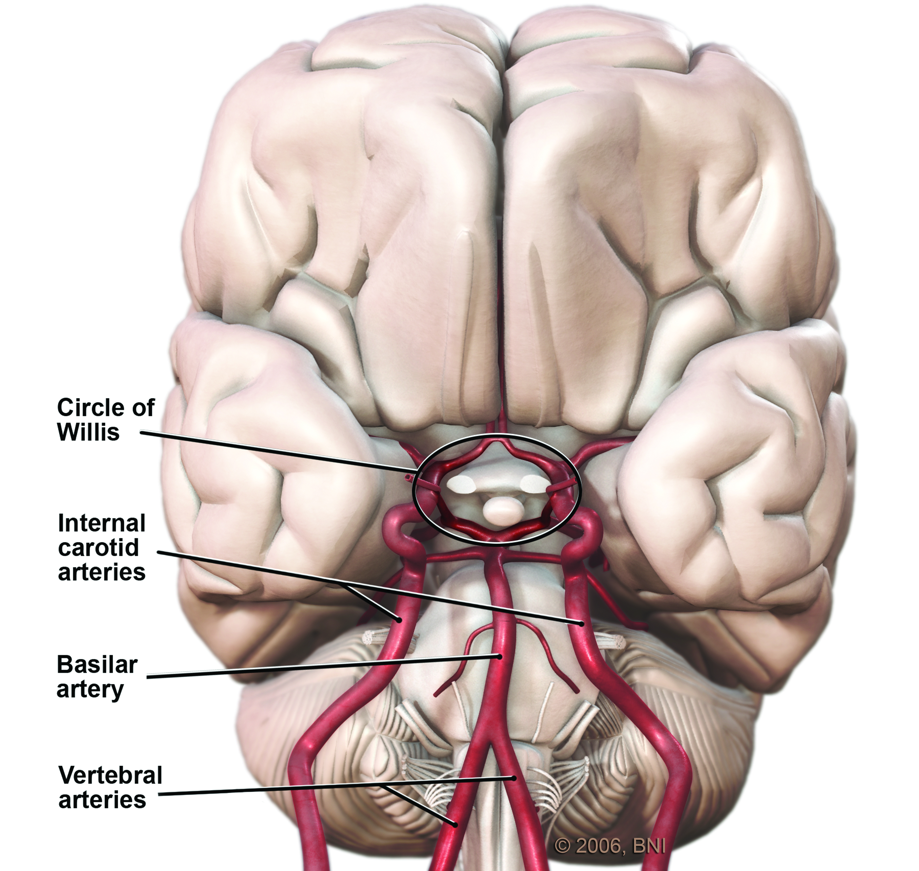
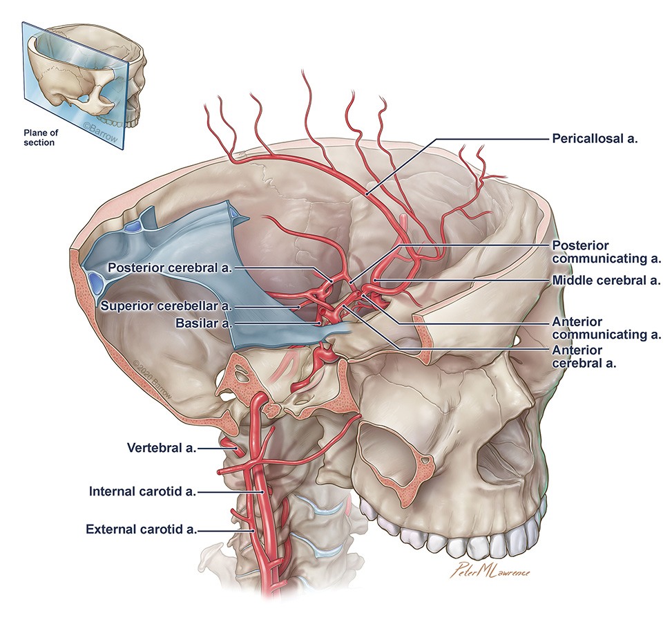
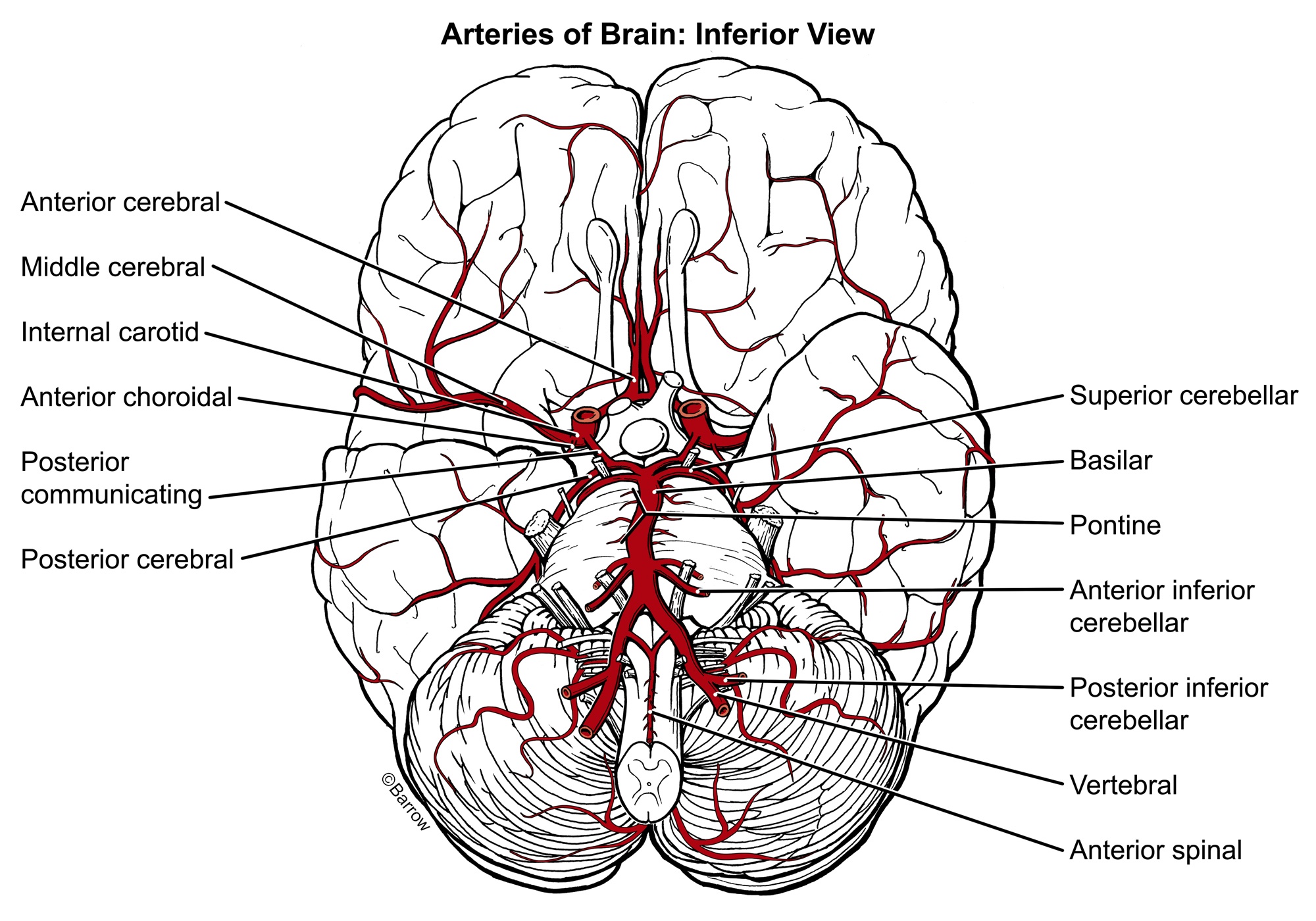
The carotid arteries are located in the neck and provide blood to the front part of the brain (known as the anterior circulation). The vertebral arteries are located at the back of the neck and provide blood to the back part of the brain (the posterior circulation). These arteries branch off into smaller blood vessels, which in turn supply different areas. Aneurysms can form on any of these arteries, though they are more common in the larger arteries at the base of the brain.
Illustrations of the Arteries at the Base of the Brain (Click to Enlarge)
Within the brain, the smallest blood vessels are called capillaries, which have thin walls that allow for the exchange of oxygen and nutrients with brain cells. The capillaries then join together to form larger blood vessels called veins, which carry the blood back to the heart to be re-oxygenated via the internal jugular veins.
The cerebrovascular system is essential for the proper functioning of the brain. Any disruption of blood flow can cause serious health problems, such as stroke or cognitive impairment. Therefore, maintaining a healthy lifestyle and managing risk factors such as high blood pressure or diabetes is important for maintaining the health of the cerebrovascular system.

Brain Aneurysm Symptoms
Cerebral aneurysms usually do not cause symptoms until they either become very large or burst. Most are found when they burst and bleed into the space between the skull and the brain. This is a serious condition called subarachnoid hemorrhage, or hemorrhagic stroke.
Symptoms of an aneurysm pressing on nearby tissue and nerves may include:
- Some growing aneurysms cause nerve compression. For instance, a posterior communicating artery aneurysm that is compressing the oculomotor nerve might cause double vision. This is a classic presentation, but also rare. Similarly, an one that is near the optic apparatus, either on the carotid artery or the anterior communicating artery, can compress the optic nerve and cause a blind spot.
- Some aneurysms that are in the basilar circulation or posterior circulation can compress the brainstem and cause symptoms such as weakness in the arms and legs, double vision, or swallowing difficulties.
- Other symptoms stemming from nerve compression can include:
- Pain above and behind the eye
- Numbness, weakness, or paralysis on one side of the face
- Dilated pupils
- Vision changes
Symptoms of a ruptured aneurysm may include:
- A sudden, severe, and explosive, or ‘thunderclap’ headache. These headaches come on in an instant and are usually described by patients as being the “worst headache of my life”, or an 11 out of 10 on the pain scale. This headache is caused by an aneurysm that has burst and started bleeding into the surrounding subarachnoid space and is a medical emergency. The subarachnoid space describes the area between the pia mater—the delicate, innermost covering of the brain—and the arachnoid mater. This space contains cerebrospinal fluid and the major blood vessels of the brain.
- Other symptoms of a ruptured aneurysm can include:
- Double vision
- Drooping eyelid
- Nausea
- Vomiting
- Stiff neck
- Sensitivity to light
- Loss of consciousness, including coma
- Seizures
Subarachnoid hemorrhage can cause serious complications, including:
- Hydrocephalus is an abnormal buildup of cerebrospinal fluid in the skull that puts pressure on brain tissue.
- Vasospasm is a narrowing of blood vessels. It reduces the amount of blood flow to the brain and can cause stroke or tissue damage.
If you are experiencing these symptoms, contact a medical professional.
Prognosis for a Ruptured Brain Aneurysm
The survival rate of a ruptured brain aneurysm used to be about 50 percent. Over time, these numbers have improved to about a 30-40% mortality rate (60-70% survival rate) as a result of advances in neurocritical care over the last 20 years.
However, roughly 25% of people who survive a ruptured brain aneurysm have cognitive deficits or discrete neurologic deficits like difficulty speaking, weakness, and other symptoms stemming from permanent brain damage.

Brain Aneurysm Treatment
Not all aneurysms bleed. Small lesions may be watched for growth and symptom onset. When deciding whether or not to treat an unruptured brain aneurysm, your doctor will consider many factors. These include the type, size, and location of the aneurysm; your health, age, and medical history; and the risks associated with treatment.
There are three kinds of treatments for brain aneurysms:
- Aneurysm clipping
- Occlusion and bypass
- Endovascular embolization
Aneurysm Clipping
Aneurysm clipping, also called microvascular clipping, is a surgical procedure that involves cutting off blood flow to the brain aneurysm. Your neurosurgeon will open up part of your skull and place a clip on the neck of the aneurysm to halt its blood supply.
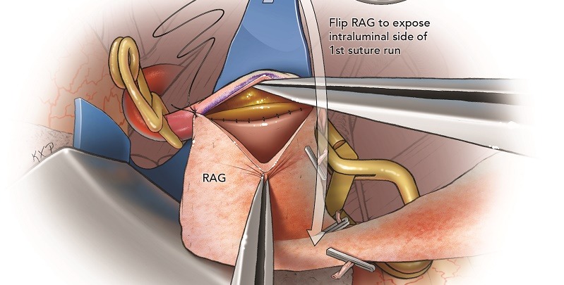
Bypass and Occlusion
Occlusion is a procedure where your neurosurgeon clamps off the entire artery leading to the brain aneurysm. This surgery is often performed when an aneurysm has damaged an artery. It may be accompanied by a bypass, in which a small blood vessel is surgically grafted to the artery. This reroutes blood flow away from the damaged section of the vessel.
Endovascular Embolization
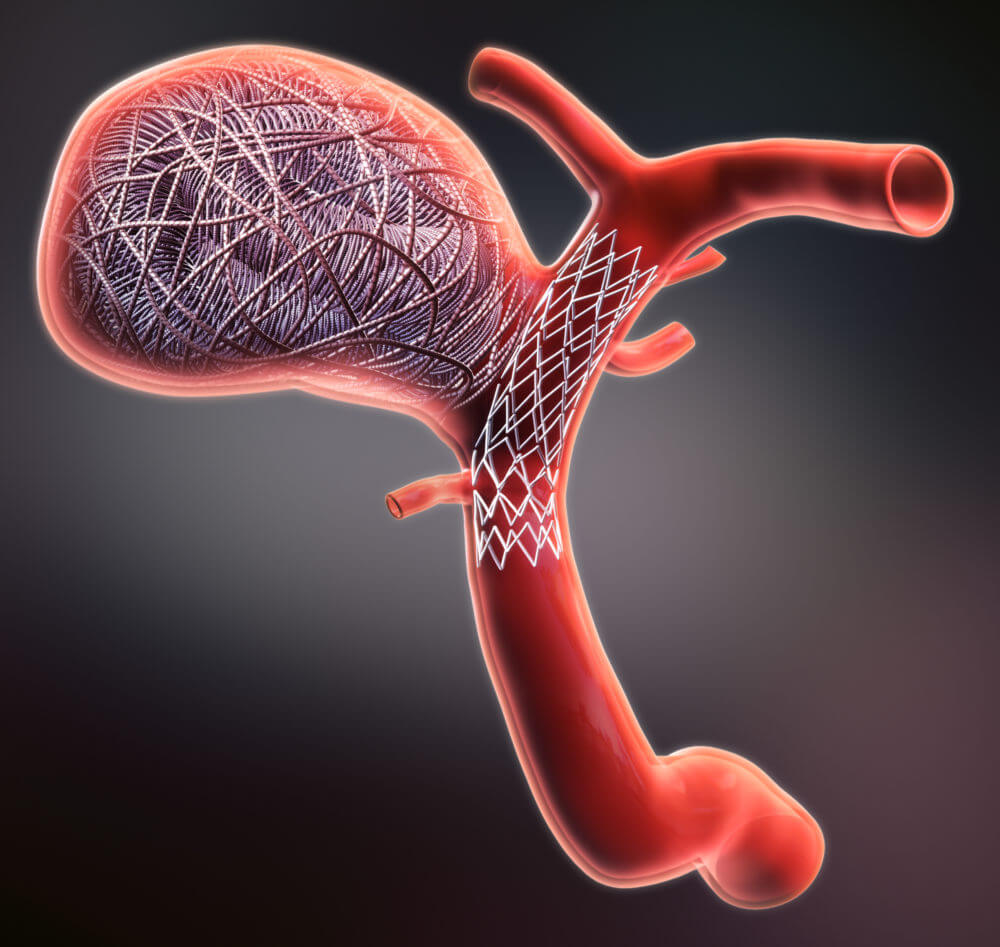
Endovascular embolization is a minimally-invasive treatment where a catheter is inserted into a major artery in the wrist or groin and threaded through the blood vessels to the site of the brain aneurysm. Coils are passed through the catheter and then released into the aneurysm.
The coils then fill the aneurysm, preventing blood from flowing through it and causing it to clot. This decreases the chances of rupture. Alternatively, an aneurysm can be treated by placing a special stent called a flow diverting stent across the base of an aneurysm. This leads to clotting and shrinkage of the aneurysm, eventually blocking it off from the main artery.
Recovery after Aneurysm Clipping or a Ruptured Brain Aneurysm
Routine, elective clipping typically requires just one night in the hospital. However, the length of stay depends on the size of the aneurysm, its location, your age, and other medical conditions you may have. Generally speaking, elective surgery requires a two-to-four week recovery time of rest and slow reintegration into your normal activities.
Recovery from an aneurysmal bleed takes much longer. It takes two weeks of in-hospital care to treat the after effects of a subarachnoid hemorrhage, which may include:
- Cerebral edema or swelling
- Increased intracranial pressure
- Vasospasm, or narrowing of the blood vessels
- Hydrocephalus
- Management of cerebrospinal fluid
Intensive care for vasospasm may include induced hypertension, angioplasty, and intra-arterial vasodilators. Patients with neurological injuries may require neuro-rehabilitation. Time to complete recovery varies greatly and depends on the severity of the hemorrhage, up to a year.
Safety of MRI after Treatment with Clips or Coils
It is safe to get an MRI after treatment with clipping or coiling. The metals used for clips or coils are MRI safe. They can, however, produce imaging artifact, or shadows, which degrade the quality of the MRI images.
Can a brain aneurysm be cured?
A cure can be achieved by excluding the aneurysm from the circulation in the brain. Indeed, the goal of surgery is always a complete cure. Clipping provides a durable cure, with recurrence rates of less than 2% over 10 years.
Endovascular therapies, such as coiling, are also intended to be curative, but have a higher recurrence rate and require retreatment in 10-15% of cases.
Patient Stories


Additional Information
Types of Brain Aneurysms
There are different types of cerebral aneurysms:
- Saccular is the most common type. It develops along weak spots in the wall of an artery.
- Dissecting forms from tears to the innermost layers of a blood vessel. It follows a traumatic injury or plaque formation.
- Mycotic is caused by a bacterial infection in the wall of an artery.
- Pseudoaneurysm is the dilatation of an artery that forms when the artery is injured by abrupt, severe trauma.
Brain Aneurysm Prevention
These lesions can occur in anyone at any age, but they are more common in adults than in children and slightly more common in women than in men. They also occur more often in people with certain genetic diseases and blood vessel disorders.
The only way to avoid a bleed is to control your risk factors, which include:
- Cigarette smoking
- High blood pressure
- High cholesterol
- Poor diet
- Lack of exercise
- Stimulant drugs like cocaine or methamphetamines
- Declining estrogen levels and menopause
You can modify these risk factors to decrease the risk of occurrence, but these measures do not prevent rupture.
The best way to prevent a bleed is early detection. If you have a family history of aneurysm, meaning that your parents or siblings have been diagnosed with a brain aneurysm or experienced a ruptured brain aneurysm, you can undergo screening using medical imaging.
How common are brain aneurysms and ruptured brain aneurysm?
About five percent of the population has at least one cerebral aneurysm. One-third of those people have more than one. Each year, these lesions bleed in 10 to 20 per 100,000 people.
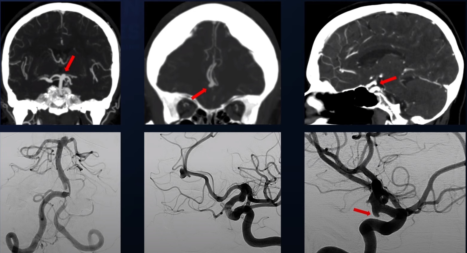
Brain Aneurysm Diagnosis
Most cerebral aneurysms go unnoticed until they bleed or are found by imaging tests done for other reasons.
The following tests may be used for diagnosis:
- Angiography
- CT scan
- MRI scan
- Lumbar puncture (spinal tap)
Can stress increase my risk?
Stress probably does not cause brain aneurysm formation, but stress can lead to a bleed. There are case reports of aneurysms that ruptured during sexual intercourse or episodes of high blood pressure.
Can exercise cause a ruptured brain aneurysm?
Strenuous exercise generally raises your heart rate and your circulation rather than your blood pressure. Some exercises that involve a lot of straining, like weightlifting, can potentially stress aneurysms.
Can you feel an aneurysm in your head?
Aneurysms can only be felt if they bleed and cause subarachnoid hemorrhage, or if they compress an adjacent nerve.
Do I need to get checked for a brain aneurysm?
You should only undergo screening for a brain aneurysm if you have a positive family history. A positive family history means that one or more of your parents or siblings have been diagnosed with a brain aneurysm.
What is the difference between a brain aneurysm and stroke?
A brain aneurysm is the anatomical sac that forms on the artery wall. If that sac bursts and bleeds into the surrounding tissue, it is known as a hemorrhagic stroke. However, hemorrhagic strokes can also be caused by high blood pressure, malformations of the arteries and veins, and aging of arteries, among other causes.
Ischemic strokes, which account for roughly 85% of all strokes, are caused by blockage of blood flow, depriving brain tissue downstream of oxygenated blood. These classic strokes result in damage or death of brain tissue.
Can a brain aneurysm change your personality?
It is unlikely for a brain aneurysm to cause personality change. Those that grow to a giant size and compress parts of the brain may affect personality, but this is exceptionally rare. Changes in personality can occur when a bleed damages parts of the brain.
Can you exercise with a brain aneurysm?
Patients with aneurysms can and should exercise. A sedentary lifestyle can contribute to poor cerebrovascular health. Many brain aneurysms are small in size (less than seven millimeters) and normal activity and exercise is encouraged for these people.
Additional Resources
Videos
Note: These videos contain surgical footage. Viewer discretion is advised.


References
- Hackett AM, Koester SW, Rhodenhiser EG, Scherschinski L, Rulney JD, Naik A, Nico E, Eberle AT, Hartke JN, Fox BM, Winkler EA, Catapano JS, Lawton MT. A comprehensive assessment of self-reported symptoms among patients harboring an unruptured intracranial aneurysm. Front Surg. 2023 Apr 21;10:1148274. doi: 10.3389/fsurg.2023.1148274. PMID: 37151867; PMCID: PMC10160638.
- Koester SW, Catapano JS, Rhodenhiser EG, Rudy RF, Winkler EA, Benner D, Cole TS, Baranoski JF, Srinivasan VM, Graffeo CS, Jha RM, Jadhav AP, Ducruet AF, Albuquerque FC, Lawton MT. Propensity-adjusted analysis of ultra-early aneurysmal subarachnoid hemorrhage treatment and patient outcomes. Acta Neurochir (Wien). 2023 Apr;165(4):993-1000. doi: 10.1007/s00701-023-05497-7. Epub 2023 Jan 27. PMID: 36702969.
- Lawton MT, Lang MJ. The future of open vascular neurosurgery: perspectives on cavernous malformations, AVMs, and bypasses for complex aneurysms. J Neurosurg. 2019 May 1;130(5):1409-1425. doi: 10.3171/2019.1.JNS182156. PMID: 31042667.
- Mascitelli JR, Mocco J, Hardigan T, Hendricks BK, Yoon JS, Yaeger KA, Kellner CP, De Leacy RA, Fifi JT, Bederson JB, Albuquerque FC, Ducruet AF, Birnbaum LA, Caron JLR, Rodriguez P, Lawton MT. Endovascular therapy versus microsurgical clipping of unruptured wide-neck aneurysms: a prospective multicenter study with propensity score analysis. J Neurosurg. 2021 Dec 24:1-8. doi: 10.3171/2021.10.JNS211942. Epub ahead of print. PMID: 34952522.