7 Journal Covers Feature Barrow Neuropub Artwork in 2021
Operative Neurosurgery, Vol. 20, Issue 3 (March)
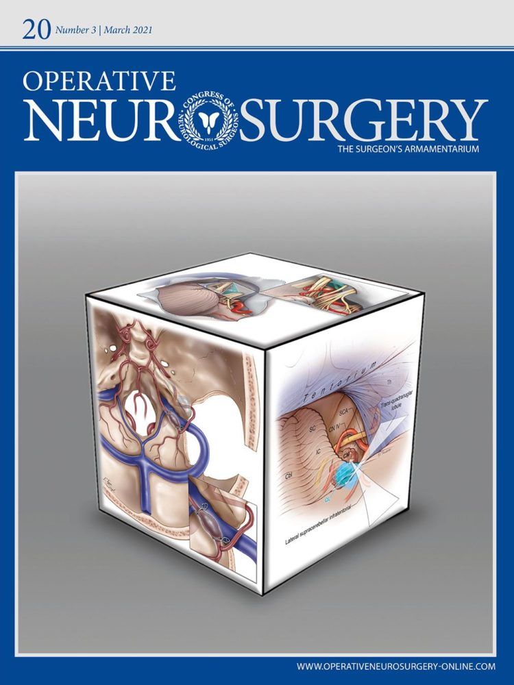
The top of the block shows the glossopharyngo-cochlear triangle (GCT), a surgical corridor located between the glossopharyngeal and cochlear nerves. Mark Schornak drew the base art for the extended retrosigmoid view, which Kristen Larson Keil expanded upon.
Journal of Neurosurgery, Vol. 134, Issue 3 (March)
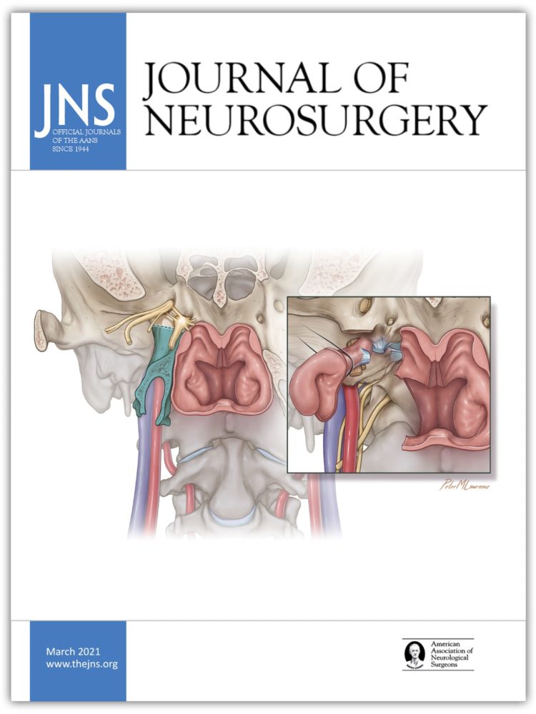
This illustration by Peter M. Lawrence shows a novel strategy for mobilizing the eustachian tube instead of resecting it while performing the endoscopic endonasal extreme medial approach to the jugular foramen and petroclival region.
Journal of Neurosurgery: Spine, Vol. 34, Issue 5 (May)
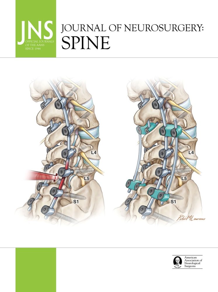
The illustration on the left depicts a traditional two-rod lumbosacral fixation construct with potentially elevated strain on posterior instrumentation (red arrow and highlighting). The illustration on the right shows the use of a supplemental rod with DTH screws to provide supplemental fixation and reduce posterior instrumentation strain. Both illustrations were created by Peter M. Lawrence.
Article: Impact of dual-headed pedicle screws on the biomechanics of lumbosacral junction multirod constructs
Journal of Neurosurgery: Spine, Vol. 35, Issue 1 (July)
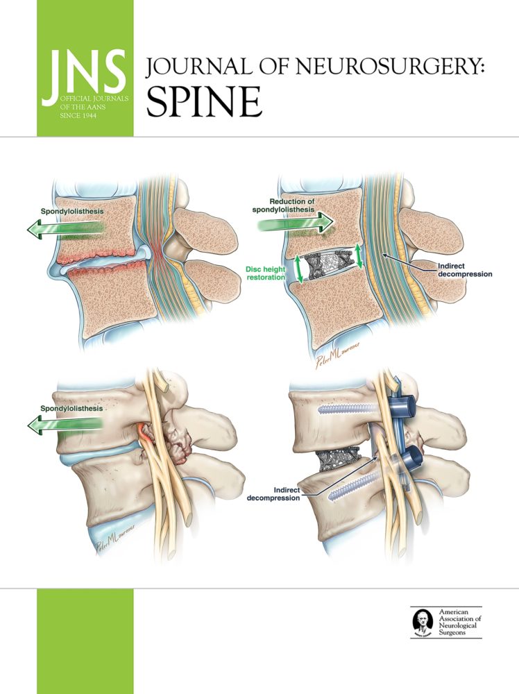
The top row features preoperative (left) and postoperative (right) illustrations showing indirect central canal decompression with reduction of the spondylolisthesis, restoration of disc height, and ligamentotaxis of the disc herniation and posterior ligamentum flavum hypertrophy. The bottom row features preoperative and postoperative sagittal illustrations demonstrating L4 nerve root impingement secondary to facet hypertrophy and degenerative changes that improve with indirect decompression of the neural foramen. All illustrations were created by Peter M. Lawrence.
Operative Neurosurgery, Vol. 21, Issue 2 (August)
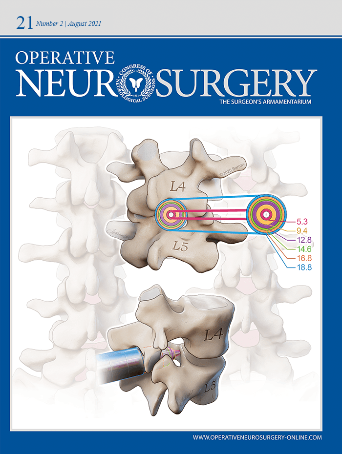
This cover illustration, created by Joshua Lai, represents the dynamic nature of the lumbar interlaminar space in extension (left) and flexion (right). In flexion, the intralaminar space can reach dimensions at which dilators of several diameters are at risk of entering the canal. The posterior oblique view (top center) demonstrates the various diameters of the dilators relative to the interlaminar space. The lateral view (bottom center) demonstrates the capacity of the lower diameter dilators to enter the canal.
Acta Neurochirurgica, Vol. 163, Issue 8 (August)
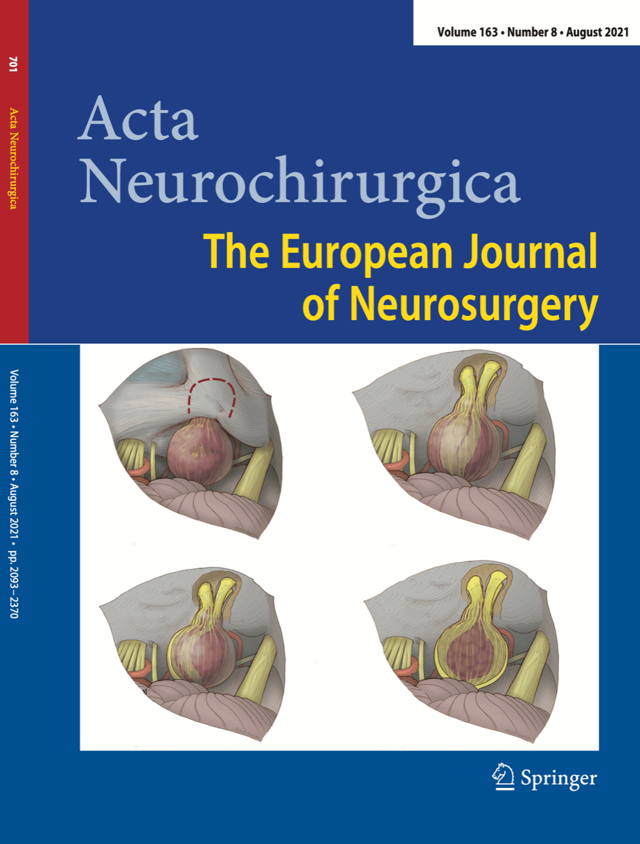
Mark Schornak created these renditions of cranial nerve configurations around a vestibular schwannoma. The top left image represents the surgical view obtained from a retrosigmoid approach for a VS resection. In the the top right image, the facial nerve is displaced anteriorly. In the bottom left image, the facial nerve is displaced inferiorly. In the bottom right image, the facial nerve is displaced superiorly and separated from the cochlear nerve by the tumor.
Journal of Neurosurgery, Vol. 135, Issue 3 (September)
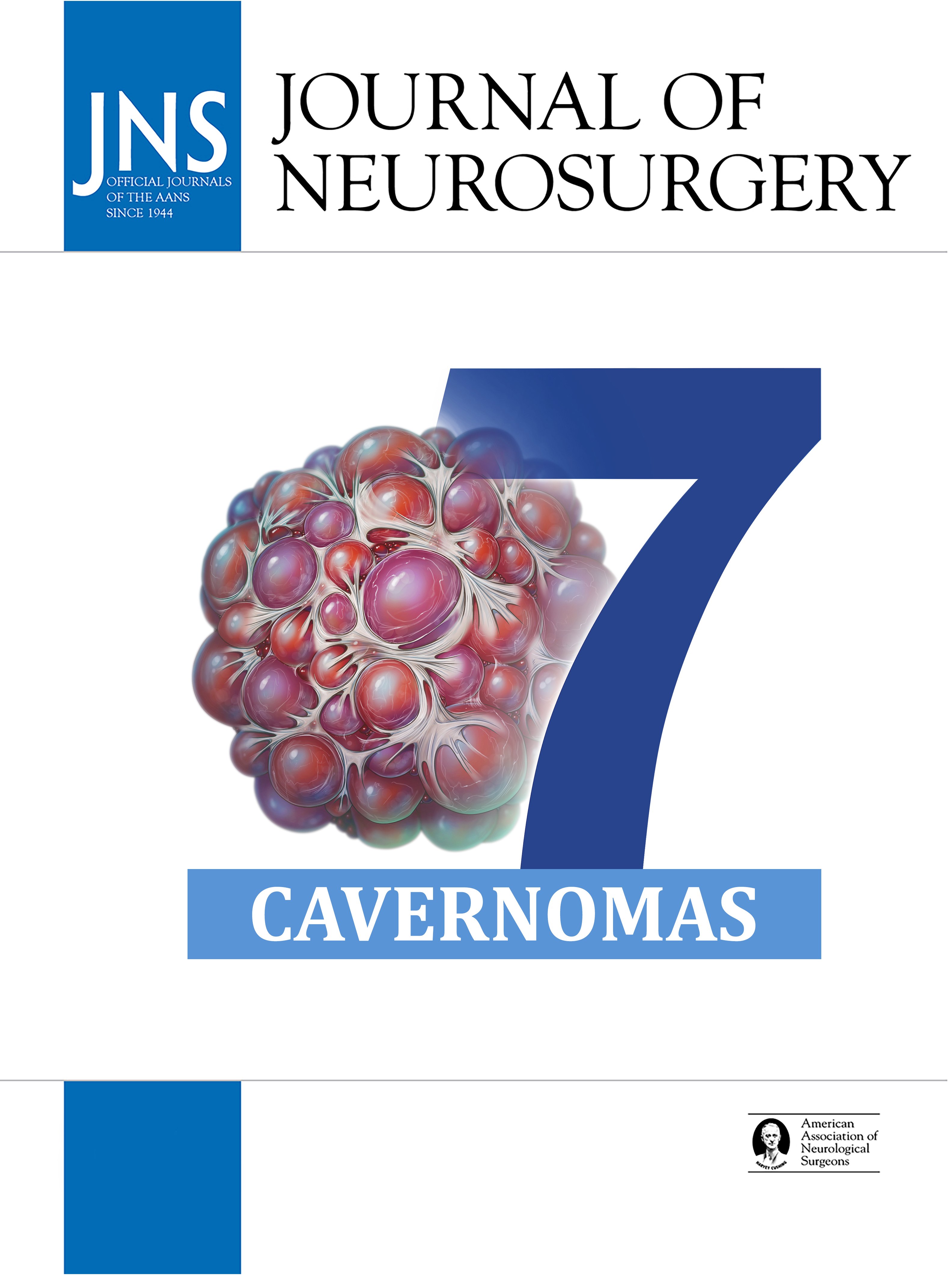
Peter M. Lawrence created this illustration of the classic mulberry appearance of a cavernoma. It represents the Seven Cavernomas series by Michael T. Lawton, MD, a collection of articles defining the tenets and techniques for the treatment of cavernous malformations, a taxonomy for classifying these lesions, and the nuances of their surgical approaches.