

Barrow-ASU Center for Preclinical Imaging
Focus and Mission
The mission of the Barrow-ASU Center for Preclinical Imaging is to provide state-of-the-art imaging technology and expertise for researchers in the Phoenix area. Our center provides a wide array of imaging modalities including MRI, bioluminescence and florescence, microPET, and microCT.
The Center is a joint effort between Barrow Neurological Institute and Arizona State University. Together, our goal is to provide essential core technologies to Valley researchers and, as a result, advance biosciences across the entire State of Arizona.
Over the past several decades, advances in imaging science have unlocked new discoveries in biology and medicine. Our facility makes this advanced technology available to researchers across Central Arizona. Using these resources, investigators can examine the in vivo anatomy and function of their preclinical models.
In addition to advanced technology, the Center for Preclinical Imaging also offers professional expertise to help researchers develop and implement their imaging protocols.
Virtual Tour

Assistant Professor, Neuroimaging Research
Featured Research
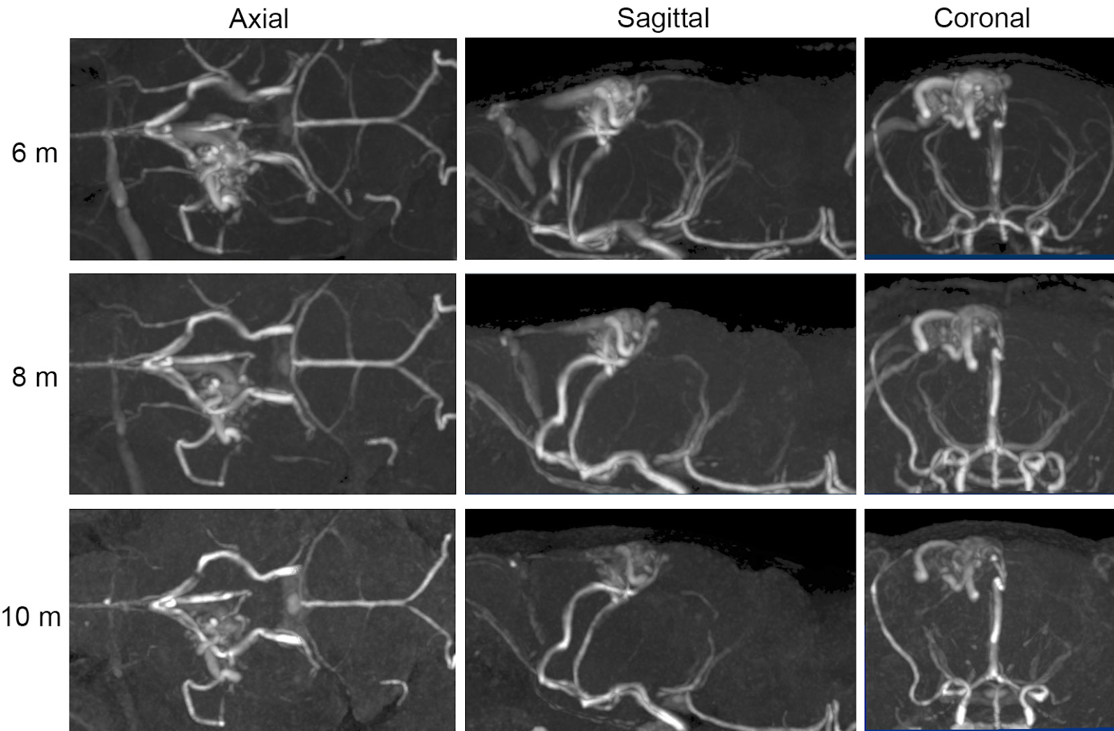
Novel experimental model of brain arteriovenous malformations using conditional Alk1 gene deletion in transgenic mice
By Chul Han, Michael J Lang, Candice L Nguyen, Ernesto Luna Melendez, Shwetal Mehta, Gregory H Turner, Michael T Lawton, S Paul Oh
Published in Journal of Neurosurgery
ABSTRACT
Hereditary hemorrhagic telangiectasia is the only condition associated with multiple inherited brain arteriovenous malformations (AVMs). Therefore, a model was developed with a genetics-based approach that conditionally deleted the causative activin receptor-like kinase 1 (Acvrl1 or Alk1) gene. Radiographic and histopathological findings were correlated, and AVM stability and hemorrhagic behavior over time were examined.
Preclinical Imaging Research
Meeting a Variety of Scanning Needs
The 7T MRI at the Center for Preclinical Imaging is equipped with a suite of imaging techniques that meet a variety of scanning needs. Some examples are as follows:
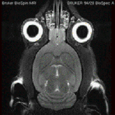
High-resolution Anatomy
The 7T has a variety of scans available for visualization of measurement of anatomy. High signal to noise and optimized protocols provide robust measurement in a variety of preclinical models.
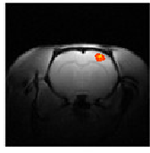
Functional MRI
Over the last decade functional MRI (fMRI) has become the most popular method for mapping brain function. Preclinical MRI provides a platform for a investigating range of topics from how pharmacologic compounds affect brain function to the better understanding of the mechanisms behind fMRI itself.
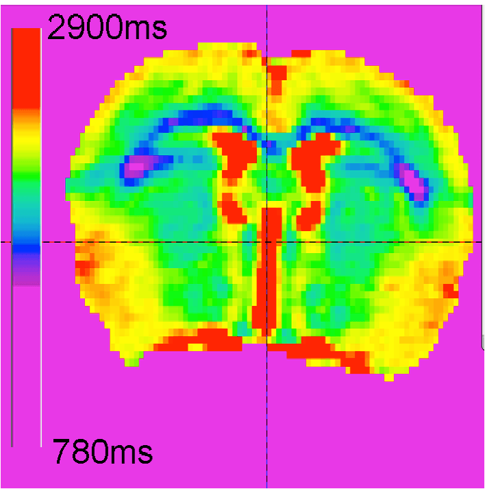
Perfusion
Measurement of perfusion and contrast agent effects are a valuable tool for examining neuropathologies such as brain tumors and stroke. Contrast agent bolus tracking, relaxivity measurements, and perfusion measurements are some of the methods available to researchers using the Center.
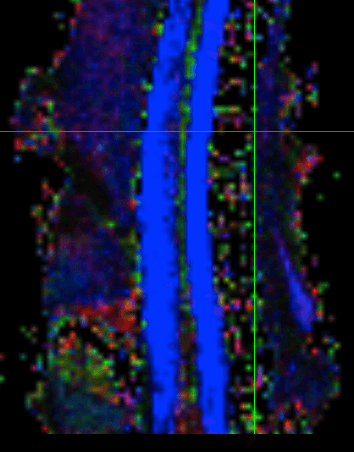
Diffusion
The 7T has routines available for acquiring images weighted for local water diffusion. Such imaging is used for characterization of disease state, such as the hypoxic edema associated with vascular stroke. Diffusion tensor imaging uses restricted diffusion data to generate images of fiber tracks in the brain or spine.
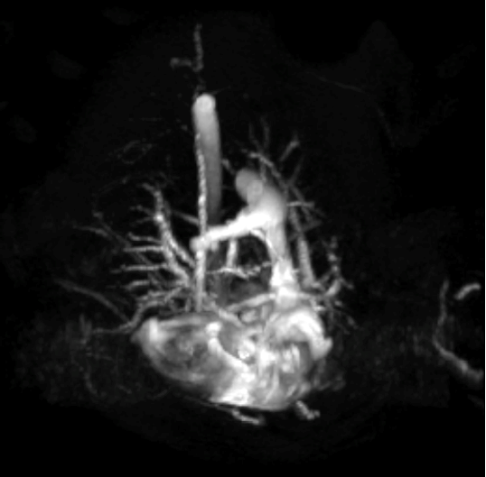
Angiography
Visualization of the vasculature is important for the study of vascular disease, stenosis, and blood flow occlusion. High-resolution, in vivo vessel imaging is possible with the use of specialized imaging techniques and/or contrast agents.

Cardiac Function
Cardiac disease is the number one cause of death in the United States. The 7T MRI at the Center for Preclinical Imaging offers state-of-the-art methods for acquiring images of the cardiac cycle to allow in vivo analysis of preclinical cardiac disease models.
Preclinical Imaging Resources
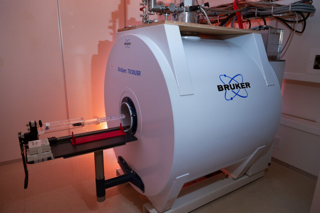
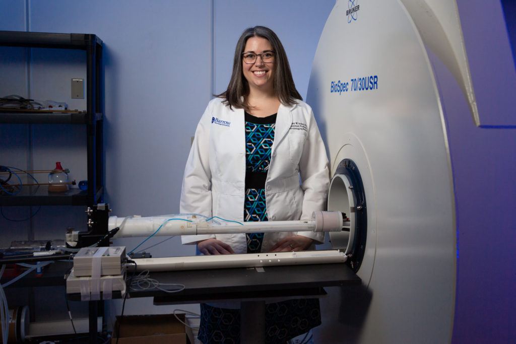
Bruker Biospec 7-Tesla MRI
This high-field magnetic resonance imaging (MRI) system has a 30-cm bore with a 200-mm gradient set (20 G/m) and a high-power 116-mm gradient insert (60 G/m). We have multiple acquisition coils to suit a variety of applications (brain, body, etc.) along with 31P and 13C coils for multinuclear spectroscopy.
The 7 Tesla (7T) MRI at the Center for Preclinical Imaging is equipped with a host of imaging capabilities to meet a variety of scanning needs:
- High-resolution anatomy. The 7T MRI has numerous scans available for visualization and measurement of anatomy. High signal-to-noise and optimized protocols allow precise measurement of anatomical structures with a spatial resolution of less than 100 microns.
- Functional MRI. Over the past decade, functional MRI (fMRI) has become the most popular method for mapping brain function. Our preclinical MRI is a platform for a range of investigations—from how pharmacologic compounds affect brain function to the quest to better understand the mechanisms underlying fMRI itself.
- Perfusion. The measurement of the effects of perfusion and contrast agents is a valuable tool for examining neuropathologies such as brain tumors and stroke. Contrast agent bolus tracking, relaxivity measurements, and perfusion measurements are some of the methods available to researchers using the Center.
- Diffusion. The 7T MRI has programmed routines for acquiring images weighted for local water diffusion. This feature can be used to characterize disease states such as hypoxic edema associated with vascular stroke. Diffusion tensor imaging uses restricted diffusion data to generate images of fiber tracts in the brain.
- Angiography. The visualization of vascular anatomy is important for the study of vascular disease, stenosis, and blood-flow occlusion. High-resolution in vivo imaging of vessels is possible with the use of specialized imaging techniques, contrast agents, or both.
- Cardiac Function. Cardiac disease is the number one cause of death in the United States. The 7T MRI at the Barrow-ASU Center for Preclinical Imaging offers cutting-edge methods for gathering images of the cardiac cycle for in vivo analysis of preclinical cardiac disease models.
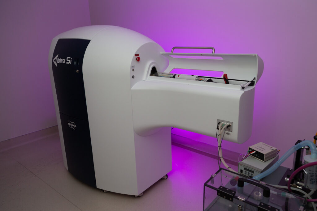
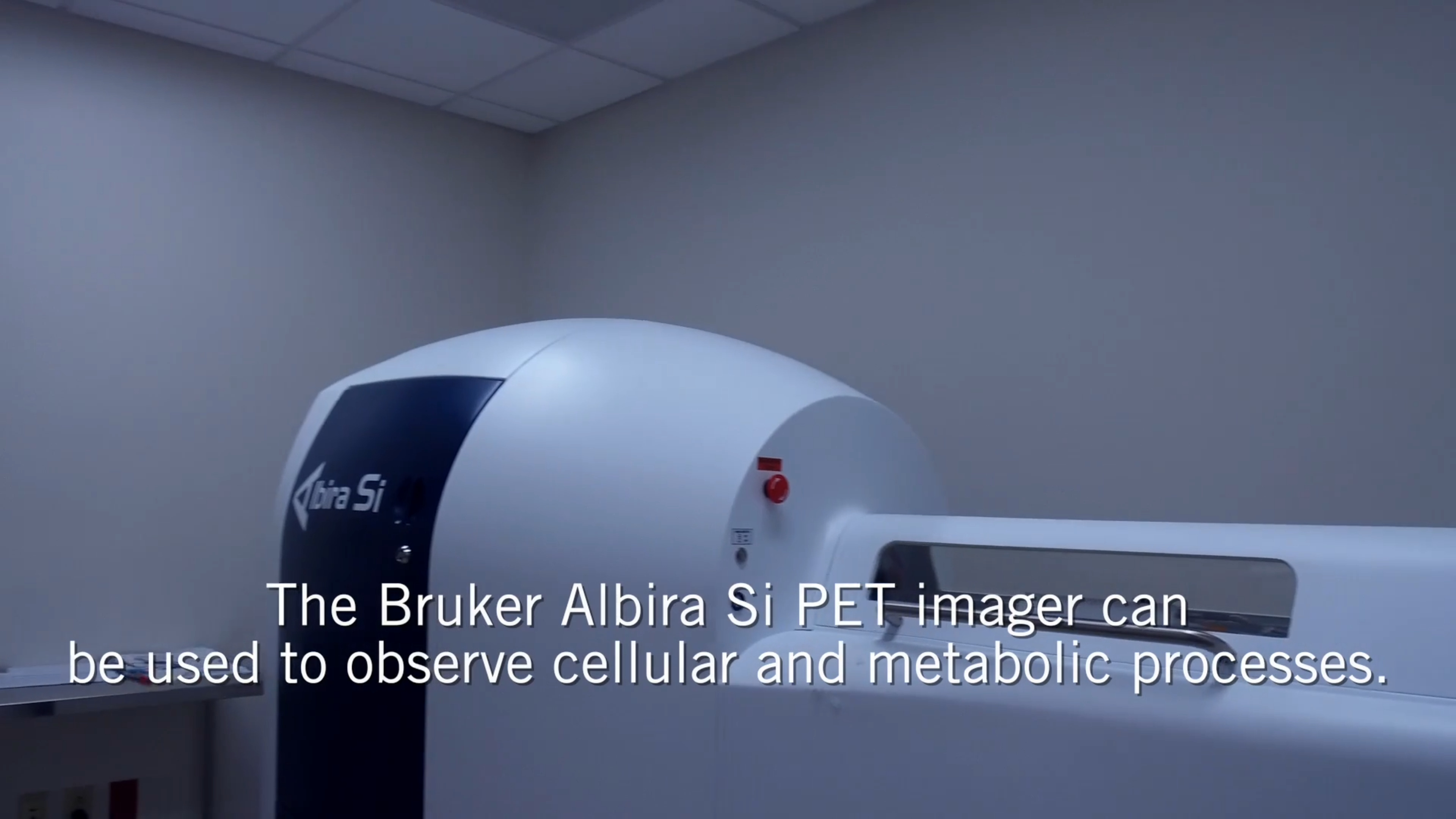
Bruker Albira Si MicroPET
Positron Emission Tomography (PET) imaging is a nuclear medicine method used to observe metabolic processes in vivo. PET uses radioactive tracers that reflect cellular and metabolic processes in the body such as glucose uptake. This imaging method is used to examine a variety of pathologies including cancer, cardiac disease, inflammation, and neurodegenerative disease.
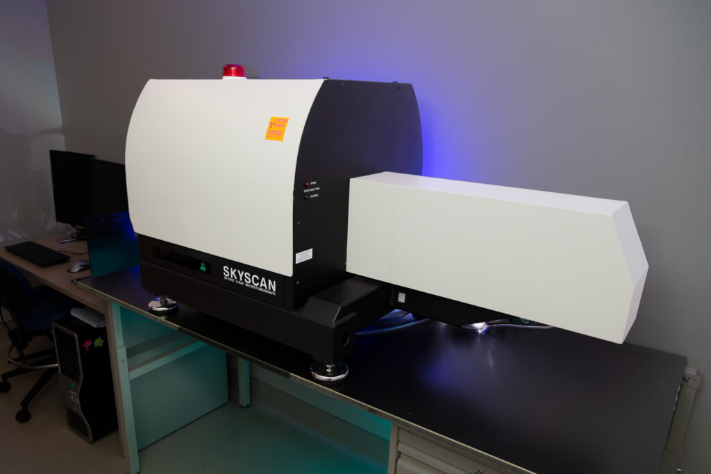
Bruker Skyscan MicroCT
Computed Tomography uses a series of X-ray images to generate renderings of cross sections of the body. It provides high-resolution images of both soft tissue and bone. It is widely used to evaluate muscle and bone disorders and study pathologies including cardiac disease, tumors, and trauma.
Xenogen IVIS Spectrum®
The Spectrum system allows noninvasive longitudinal measurement of disease states, cell trafficking, and gene expression patterns. Both fluorescent and bioluminescent reporters can be imaged with this system as well as with 2- and 3-dimensional tomography.
Procedure Rooms
Two procedure rooms equipped with medical gasses, fume hoods, and basic laboratory equipment (e.g., scales, pipettes) are available for study preparation. Holding rooms are also available for a per diem fee (Contact Us).
Computing
A data analysis workstation is available for end users to work with their data offline. A file-transfer protocol (FTP) server also is available to facilitate remote access to study data and data transfer to an off-site location.
Application for Use of CPI Resources
How can we help you?
Ashley Stokes, PhD
Program Director, Preclinical Imaging
Barrow Neurological Institute
St. Joseph’s Hospital and Medical Center
- Phone: (602) 406-8789
- Fax: (602) 406-3911
- E-mail: Ashley.Stokes@Barrowneuro.org
Core Staff Members
Ashley Stokes, PhD
Program Director, Barrow Neurological Institute
Phone: (602) 406-8789
E-mail: Ashley.Stokes@BarrowNeuro.org
Alberto Fuentes
Research Engineer II
Phone: (602) 406-5595
E-mail: Alberto.Fuentes@CommonSpirit.org
Affiliated Researchers
Vikram Kodibagkar, PhD
Associate Professor, Arizona State University
E-mail: Vikram.Kodibagkar@asu.edu

Call Us
Ashley Stokes, PhD
(602) 406-5663