Pioneering Device May Advance Neurosurgery, Researchers Say
Barrow Neurological Institute, University of Bern, and C. Besta Neurological Institute researchers collaborate to develop new imaging technology
Researchers at Phoenix’s Barrow Neurological Institute, the University of Bern in Switzerland, and the C. Besta Neurological Institute in Italy have used a hand-held microscope that provides neurosurgeons a picture of malignant brain tumor cells in real-time as they remove them. The study was recently published in the Journal of Neurosurgery.
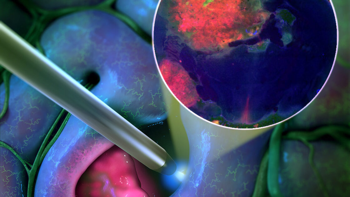
The new tool – Confocal Laser Endomicroscopy – CONVIVO by Zeiss Medical Technology – is the size of a pen and is the first hand-held microscope that provides images of tissue at cellular resolution. The pioneering device uses fluorescence to illuminate the malignant glioma cells. Combined with existing imaging technology, neurosurgeons are exploring the use of the technology to achieve an improved assessment of the indistinct tumor margin and a more extensive or optimal tumor resection than they could using existing technology.
“This unique innovation is advancing neurosurgery on a cellular basis,” said Mark Preul, MD, director of neurosurgery research at Barrow Neurological Institute, part of Dignity Health St. Joseph’s Hospital and Medical Center. “We have shined a new light on brain tumor operations.”
An imaging technology that is thought to be reliable in the neurosurgical operating room for removal of malignant gliomas relies on giving patients a drug orally before surgery. The drug (5-ALA) makes the tumor cells glow under certain fluorescent light conditions as the surgeon operates. However, there are problems with this technology because the tumor cells do not always fluoresce as expected, even though they are malignant; and malignant glioma cells fluoresce variably because they may have different genetics or metabolism. Very importantly, at the margin of these malignant tumors, the tumor cells do not glow much or lose their glow under intense microscope illumination.
By using this new microscope that uses a different fluorescent drug in addition to the 5-ALA drug, the neurosurgeon can better discriminate the margin region of these invading tumors and identify remaining tumor cells that are at the invading tumor margin.
“Using the two fluorescent systems together appears to provide a significant advantage to better identify the tumor margin and invading tumor cells,” Dr. Preul said.
Confocal Laser Endomicroscopy – CONVIVO was pioneered first for neurosurgery in clinical trials at Barrow Neurological Institute with Zeiss and shows the tissue and cells in real-time on the fly to the neurosurgeon and can be used in a secure image communication system that connects the neurosurgeon in the operating room with the neuropathologist. While the neurosurgeon scans and images the tissue, the neuropathologist can be anywhere while viewing the same images and discussing with the neurosurgeon.
“This unique innovation is advancing neurosurgery on a cellular basis. We have shined a new light on brain tumor operations.”
Mark Preul, MD, Director of Neurosurgery Research
“In the short term, with this hand-held cellular imaging technology, neurosurgeons can begin to better detect the difficult-to-define margin regions of malignant glioma and identify cellular areas that could be resected further, thereby optimizing the extent of resection of these tumors,” Dr. Preul said. “While long-term implications are yet to be seen, we expect that extending the resection margin for these tumors or identifying leftover tumor tissue for resection could improve the effectiveness of post-operative therapies and result in longer survival for patients.”

