Minimally Invasive Lateral Thoracic Discectomy
Overview of Minimally Invasive Lateral Thoracic Discectomy
Minimally invasive lateral thoracic discectomy (or mini-open lateral thoracic discectomy) is a procedure in which a herniated thoracic intervertebral disc is surgically removed from the spinal column. Intervertebral discs separate the bones of the spine, called vertebrae. They also act as shock absorbers and enable movement.
Traditionally, spinal surgeons performed thoracic discectomies through an anterior (front) or posterior (back) approach. Our spinal neurosurgeons helped to develop the minimally invasive lateral approach, in which they access the spine through a small incision in the side of the body.
The benefits of this approach include:
- A shorter hospital stay
- Less post-operative pain
- Reduced blood loss
- Reduced risk of complications
- Usually no need for chest tubes
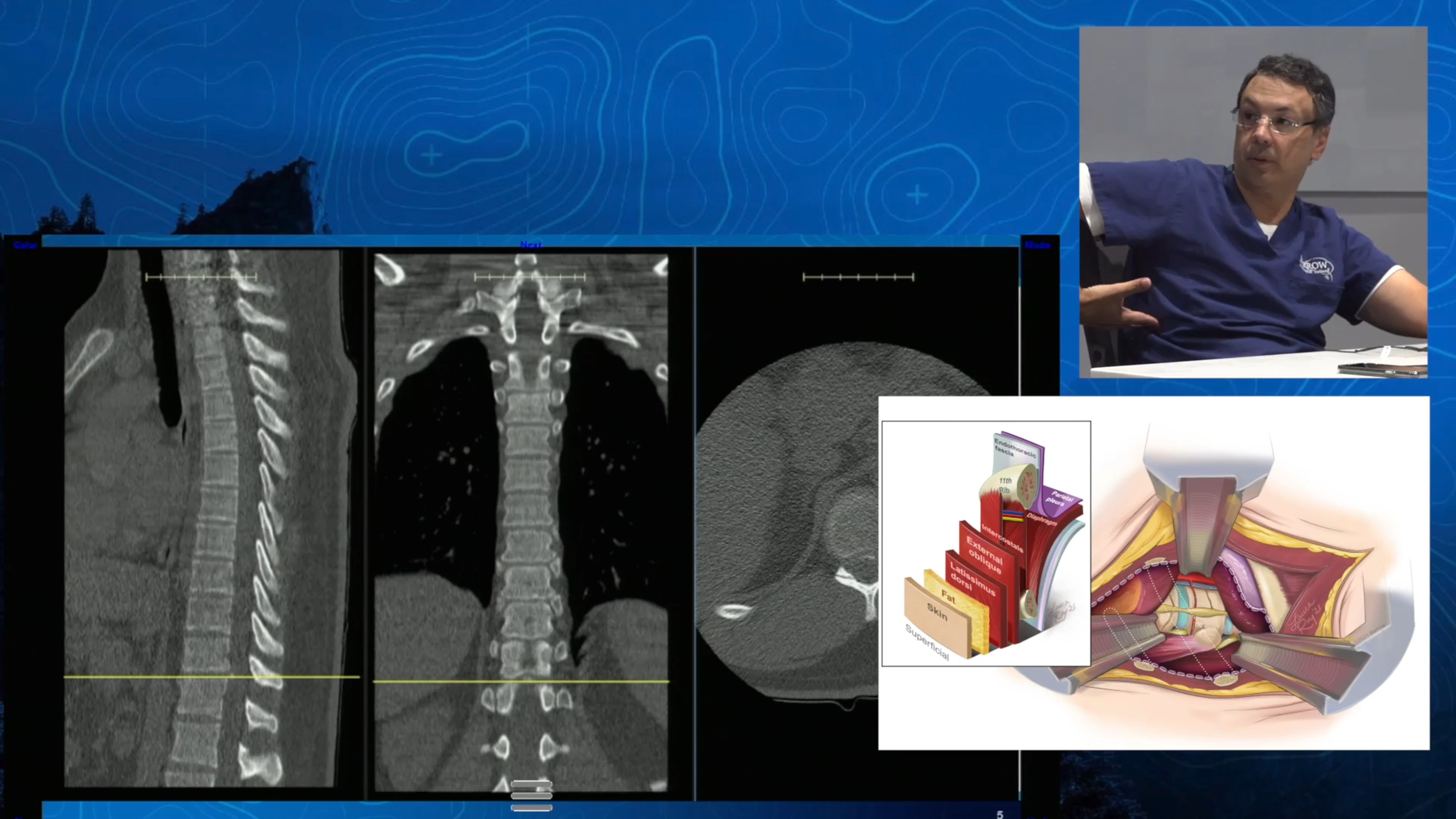
In the lateral approach, also known as the retropleural approach, the patient lies on their side. The spine surgeon makes a small incision over the rib cage and may remove part of a rib. Specialized instruments, advanced microscopes, and navigation techniques allow the spine surgeon to work through the small incision. Neurophysiological monitoring reduces the risk of spinal cord injury during surgery. To access the spine, the spine surgeon moves the tissue that covers the lungs (the pleura). Sometimes deposits of calcium can form in the disc space—a process called calcification. If the herniated thoracic disc is calcified, the spine surgeon will drill the hardened disc until it is paper-thin before removing it.

What is lateral thoracic discectomy used for?
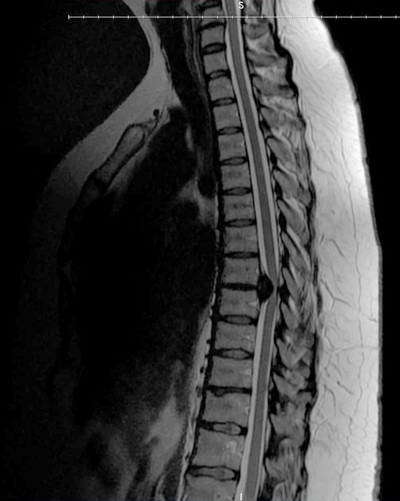
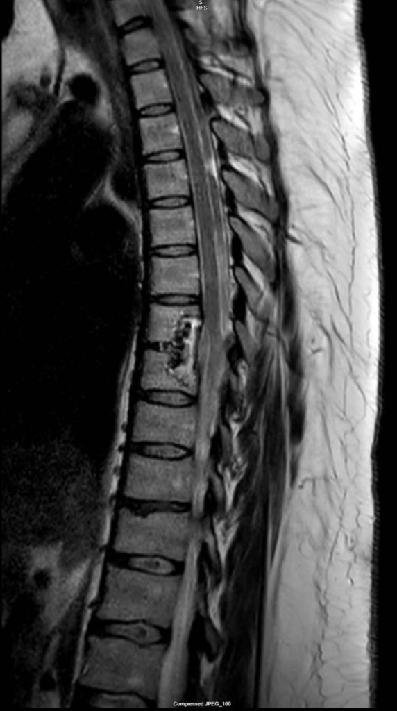
An MRI showing a calcified herniated thoracic disc compressing the spinal cord.
Minimally invasive lateral thoracic discectomy is used to treat thoracic disc herniation. This is a spinal condition in which the soft center of an intervertebral disc (the nucleus pulposus) pushes through a tear in the outer layer of the disc (the annulus fibrosus) and into the spinal canal.
Discs can tear due to degeneration, injury, or a combination of both. The protruding disc fragment can put pressure on the spinal cord or the nerves entering and exiting it, causing upper back and chest pain and spinal cord dysfunction.
Herniated discs are much more common in the lumbar and cervical regions of the spine. The thoracic region, which is the largest segment of the spinal column, is the least mobile region and therefore the least susceptible to disc herniation. However, thoracic disc herniations may be underdiagnosed because symptoms can be subtle and unspecific.
Herniated discs in the thoracic region have a tendency to become calcified, also known as hard disc herniation.
Am I a good candidate for minimally invasive lateral thoracic discectomy surgery?
You may be a good candidate for minimally invasive lateral thoracic discectomy if you have a thoracic disc herniation that is significantly affecting your quality of life or compressing your spinal cord.
Discectomies in the thoracic region of the spine are more complicated and carry an increased risk of spinal cord injury compared to discectomies in the lumbar or cervical regions. Our spine surgeons carefully evaluate each patient before making treatment recommendations.
Most cases of thoracic disc herniation can be managed with a more conservative approach consisting of rest, anti-inflammatory medication, and physical therapy.
Thoracic disc herniations that are not producing symptoms, which are found when someone has imaging tests done for an unrelated problem, can be monitored with imaging over time.
You may not be a good candidate for this procedure if you have other health conditions, such as severe obesity or heart problems.
Lateral Thoracic Discectomy Video