Colloid Cyst
Overview
Colloid cysts are brain lesions filled with a thick, gel-like substance called colloid. They are usually found on the roof of the third ventricle, a cavity in the center of the brain where cerebrospinal fluid is produced and flows through on its way to the outside of the brain. This fluid cushions the brain and spinal cord.
These cysts are usually benign and may sit quietly in the brain for years, but they can eventually grow larger and cause neurological symptoms. Colloid cysts can obstruct the flow of cerebrospinal fluid, causing fluid buildup in the ventricles of the brain (hydrocephalus) and increased pressure within the skull. This pressure can cause headaches, nausea, vomiting, imbalance, confusion and, in rare cases of complete obstruction of the spinal fluid, result in brain herniation (downward displacement of the brain resulting in the blockage of blood vessels to the brain) and sudden death.

Colloid Cyst Symptoms
Symptoms of a colloid cyst depend on its size but may include:
- Headaches
- Nausea
- Vomiting
- Confusion
- Difficulty walking
- Imbalance
- Brief loss of consciousness
Colloid Cyst Treatments
If the cyst is not causing symptoms, your doctor may recommend observation only. If the cyst appears to be at risk of obstructing the flow of cerebrospinal fluid, or if it is causing increased pressure within your skull, surgery is necessary.
Surgical Treatment for Colloid Cysts
Options for the surgical treatment of a colloid cyst include placement of a shunt, which may be used to bypass the obstruction of cerebrospinal fluid and divert it into your abdomen.
Direct removal of the cyst by surgery is usually preferred since it relieves the obstruction and avoids the dependency on the shunt and the possible risks of shunt infections or failures. Complete surgical removal of colloid cysts is challenging because of where they are located in the brain; however, modern techniques, such as image guidance, have increased the safety and efficacy of this option.
Damage to short-term memory function is the main risk of surgical removal, since the cysts are located next to the fiber tracts in the brain responsible for storing new memories. To access the cyst with minimal risk to short-term memory, your surgeon may use a small open surgical approach between the cerebral hemispheres with the operating microscope, or use an endoscopic approach depending on the location and size of your cyst.
Individualized Care
There is no single recipe or “cookbook” approach that works best for everyone with a brain tumor. Every brain tumor is unique, as is each patient. Personalized medicine approaches, such as tumor profiling to look for specific gene mutations, can help determine the best therapies available for you.
Common Questions
How common are colloid cysts?
Colloid cysts are rare. They account for an estimated 0.5 to one percent of all primary brain tumors.
Who gets colloid cysts?
Colloid cysts are believed to form during embryonic development of the central nervous system. However, symptoms are not usually present until adulthood.
How are colloid cysts diagnosed?
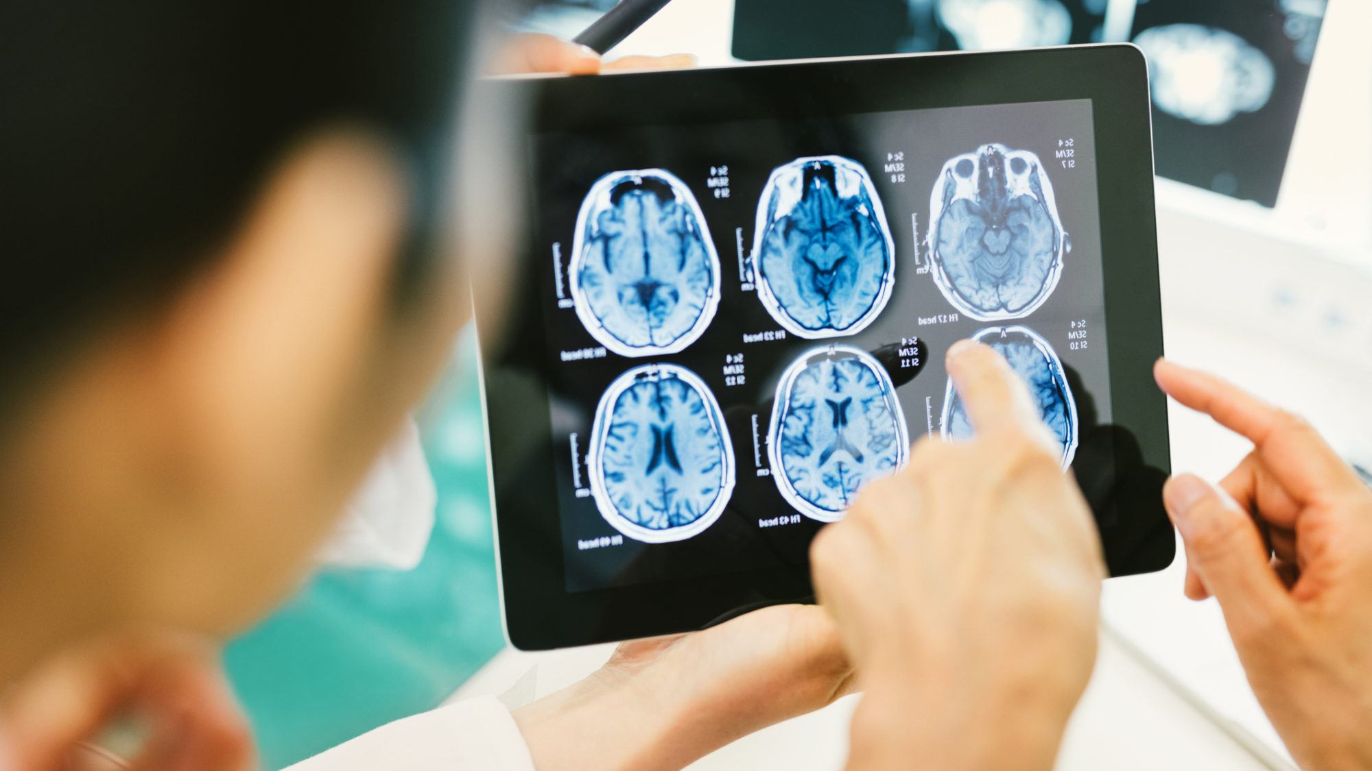
Imaging tests, such as computed tomography (CT) and magnetic resonance imaging (MRI) scans, can be used to diagnose colloid cysts.


