
Congress of Lateral Skull Base Surgery Temporal Bone Course
5th Annual Congress of Lateral Skull Base Surgery
Course Description
The temporal bone constitutes one of the most anatomically complex locations in the human body. For this reason, a comprehensive knowledge of the complex three-dimensional anatomy of this region is vital for achieving successful surgical outcomes. The principal aim of the annual congress is to provide comprehensive cognitive and skills training for the safe and efficient completion of lateral skull base approaches. After a successful inaugural session in May 2018, this year’s course will be the 5th iteration of the congress!
Seats will be reserved for an international audience of senior neurosurgical residents, neurosurgical skull base fellows, neurotology fellows, and practicing surgeons/faculty. Attendees will benefit from a concise lecture series by Barrow experts and renowned guest faculty, extensive time in the dissection lab working on preserved cadaveric whole heads, use of clinical-grade operative microscopes, and direct one- on-one mentorship.
This year, we are pleased to welcome two guest faculty representing ENT/Neurotology (Tom Roland). In addition to offering our standard training regimen on traditional lateral skull base approaches, we will welcome Dr. Roland to help provide didactics and skills training on special topics in lateral skull base surgery. The focus of the fifth congress will be on auditory brainstem implantation and advanced combined petrosal approaches.
Course Participants
- Senior neurosurgical residents
- Neurosurgical skull base fellows
- Neurotology (ENT) fellows
The course will be limited to 16 individuals; two per station, 8 stations, plus one pro-section.
Course Learning Objectives
At the conclusion of this course, participants should be able to:
- Describe all major anatomical structures of the temporal bone and petrous apex from four perspectives (posterior fossa, middle fossa, lateral surface structures, intra-temporal)
- Perform lateral skull base surgical approaches and identify key visual-spatial relationships pertinent to the treatment of neoplastic, neurovascular, and infectious disorders
- Demonstrate surgical proficiency operating under high powered microscopy within the confines of the temporal bone and in using various otologic micro- instrumentation
- Identify key anatomical landmarks identifying the lateral recess of the 4th ventricle and successfully place an auditory brainstem Implant paddle electrode
- List the key steps to achieve a successful implantation and activation of an auditory brainstem implant
Registration Information
| Attendee Type | price |
|---|---|
| Residents & Fellows | $200 |
| Non-trainee | $2,500* |
*Non-trainee slots will be made available on March 8, 2024 if the course has not filled
Registration fee includes: Symposium, program materials, and continuing education credits.
Refund and cancellation policy: To ensure adequate spaces and planning for the course, no refund are given for canceled reservations.
In-Person
April 12-13, 2024
Phoenix, Arizona

Date, Time, and Location
In Person
Friday & Saturday, April 12-13, 2024
Loyal and Edith Davis Neurosurgery Research Laboratory
Barrow Neurological Institute
350 West Thomas Road
Phoenix, Arizona 85013
Speaker Information
All times refer to Arizona time (Pacific Daylight Time).
| SPeaker | Topic |
|---|---|
| Friday, April 12 – Arrival and Registration | 7:00 AM |
| Robert F. Spetzler, MD | Course Intro, Overview, and Announcements (7:20 AM) |
| Shawn Stevens, MD | Anatomy of the Temporal Bone (7:30 AM) |
| Tom Roland | Guest Faculty Grand Rounds No. 1 (8:00 AM) |
| TBD | Guest Faculty Grand Rounds No. 2 (8:45 AM) |
| Transition to Dissection Lab | 9:30 AM |
| Shawn Stevens, MD | Trans-Temporal Approaches – Technique, Pearls, and Pitfalls (9:45 AM) |
| Shawn Stevens, MD | Dissection Stations; Part 1 Embalmed whole head, RIGHT side Dissection Focus: Trans-labyrinthine, Trans-Otic Approach (10:00 AM) |
| Lunch | 12:45 PM |
| Transition to Dissection Lab | 1:25 PM |
| Tom Roland | Infratemporal Fossa Approach – Basics of ABI (1:40 PM) |
| Tom Roland | Dissection Stations; Part 2 Embalmed Whole Head, LEFT Side Dissection Focus: Middle Fossa, Extended Petrosal, and Combined Approaches (2:00 PM) |
| Tom Roland Shawn Stevens, MD Kaith Almefty, MD Randall W. Porter, MD Kris A. Smith, MD | End of Day Discussion: Ask the Experts Panel (5:00 PM) |
| Adjourn | 5:30 PM |
| Course Participant and Faculty Dinner. Dress Casual. Location TBD. | 6:30 PM |
| Saturday, April 13 | Breakfast Reception (7:00 AM) |
| Shawn Stevens, MD Kaith Almefty, MD | Sat Overview. Middle Fossa and Combined Petrosal (7:30 AM) |
| Transition to Dissection Lab | 8:30 AM |
| Kaith Almefty, MD | Dissection Stations; Part 3 Embalmed Whole Head, LEFT Side Middle Fossa Approaches, Petrous Apex, Cavernous Sinus (8:45 AM) |
| Lunch | 12:00 PM |
| Transition to Dissection Lab | 12:45 PM |
| Various Sponsors | Sponsor Stations Available Through Afternoon Session Sponsor 1: Intro to the ABI, Technology Overview | Cochlear Corp. Sponsor 2: Advances in Surgical Instrumentation | Stryker |
| TBD | Posterior and Combined Petrosal Approaches (1:00 PM) |
| TBD | Dissection Stations; Part 4 Embalmed Whole Head, LEFT Sides Dissection Goals: Complete Prior Dissections, Posterior and Combined Petrosal Approaches (1:20 PM) |
| Kaith Almefty, MD Shawn Stevens, MD | Final Words and Adjournment (5:00 PM) |
Laboratory Contact Information
Research Laboratory: (602) 406-3268
Fax: (602) 406-4153
Email: William.Bichard@CommonSpirit.org
Barrow Neurosurgery Research Laboratory
Marion Rochelle Neuroscience Research Center
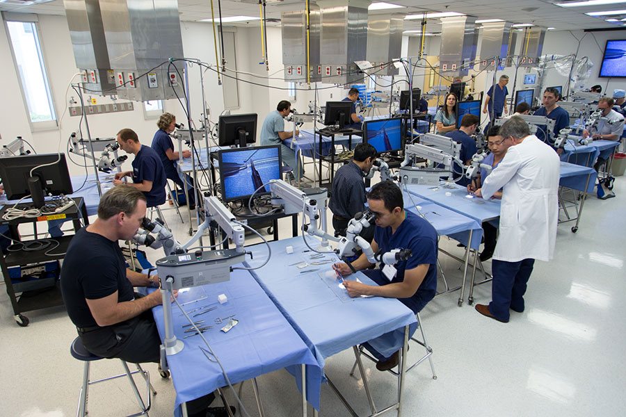
The course will take place at the Neurosurgery Research Laboratory of the Department of Neurosurgery at Barrow Neurological Institute, which is a world-class education, training, and research facility with a specialization in neurosurgical anatomy. The facility is well known for exquisite cadaver tissue specimens and features independent surgical stations fully equipped with operating microscopes, suction, irrigation, standard head frames, microsurgical and power instrumentation, 3D surgical projection, high definition flat screens, and fully-trained attendant staff.