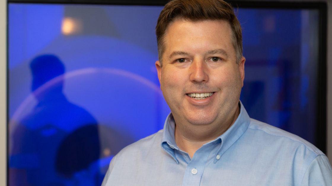
Dr. Richard Dortch: #TheMindBehind Imaging Research for Nerve Degeneration, Repair
As an undergraduate student majoring in electrical engineering, Dr. Richard Dortch envisioned a career at either a communications or utility company. But while working on cochlear implants for a school project, he began to consider a different path.
“I saw that some engineering methods that we were developing could possibly have real-world impacts on people with various neurological conditions,” he recalled.
After graduating with his bachelor’s degree in electrical engineering from the University of Tennessee at Chattanooga, he went on to study biomedical engineering. He earned both a master’s degree and PhD from Vanderbilt University in Nashville.
Dr. Dortch also completed his postdoctoral training at Vanderbilt. He then joined the faculty as an assistant professor in the departments of Radiology and Biomedical Engineering.
“The idea of taking the technical skills, the things I love to do, and then actually apply them to impact patient care is extremely rewarding,” he said.
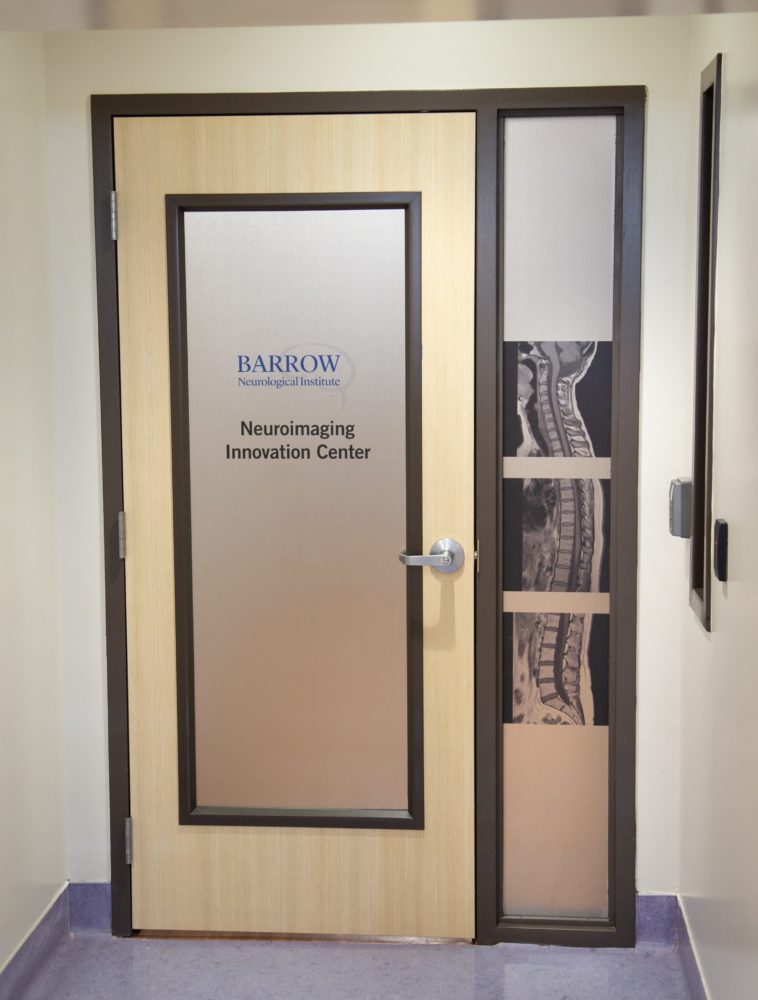
Dr. Dortch had long admired the translational research taking place at Barrow Neurological Institute in Phoenix, where his Vanderbilt colleague Chad Quarles, PhD had landed in 2015. Therefore, Dr. Dortch decided to apply for a faculty position. He joined the Barrow team as an associate professor in October 2019, establishing his own laboratory in the Neuroimaging Innovation Center.
Since then, Dr. Dortch has collaborated with neurosurgeons, neurologists, and fellow imaging scientists at Barrow to develop innovative imaging technologies. The goal of these new methods is to help guide surgery, improve diagnostics, and measure both disease progression and how patients are responding to therapies.
His lab primarily focuses on peripheral nerve and spinal cord imaging. He uses magnetic resonance imaging (MRI) techniques to gather information on the degeneration and repair of nerve structures.
R21 Grant: Exploring MRI Biomarkers for Inherited Neuropathies
One of Dr. Dortch’s current projects focuses on Charcot-Marie-Tooth (CMT) disease, a type of inherited neuropathy. This rare disease damages the peripheral nerves, which are the nerves that extend from the spinal cord to the rest of the body. CMT affects motor nerves, causing muscle weakness and deterioration. It also affects sensory nerves, inhibiting a person’s ability to feel stimuli such as temperature and touch.
Scientists have made strides toward a treatment for the disease. However, the lack of sensitive and objective biomarkers has hindered their ability to see if experimental treatments are effective. Biomarkers are measurable indicators of a disease, like blood sugar levels in diabetes.
“Disease progression is slow in patients with CMT, so we need markers that are more sensitive to progression and hopefully to treatment response,” Dr. Dortch explained.
He and Jun Li, MD, PhD, at Wayne State University in Detroit applied for a grant to study whether their nerve MRI methods might offer a solution.
In April, their proposal received a competitive R21 grant of $440,344 in total funding from the National Center for Advancing Translational Sciences at the National Institutes of Health (NIH). R21 grants are exploratory, meaning they provide support for early and conceptual stages of projects.
“The idea is that we’ll establish the methods in these two years,” Dr. Dortch said. “If successful, we can apply for a larger grant that would allow us to push these methods out to additional sites and test them in larger clinical trials,” Dr. Dortch said.
Accelerating MRI Research in MS Through Multi-Site Collaboration
Dr. Dortch is also working to develop MRI biomarkers for multiple sclerosis. In this disease, the body’s immune system mistakenly attacks the myelin sheath that surrounds and protects central nerves.
Existing treatments don’t promote myelin repair, an avenue that Dr. Dortch says is impeded in part by a lack of reproducible myelin-specific biomarkers.
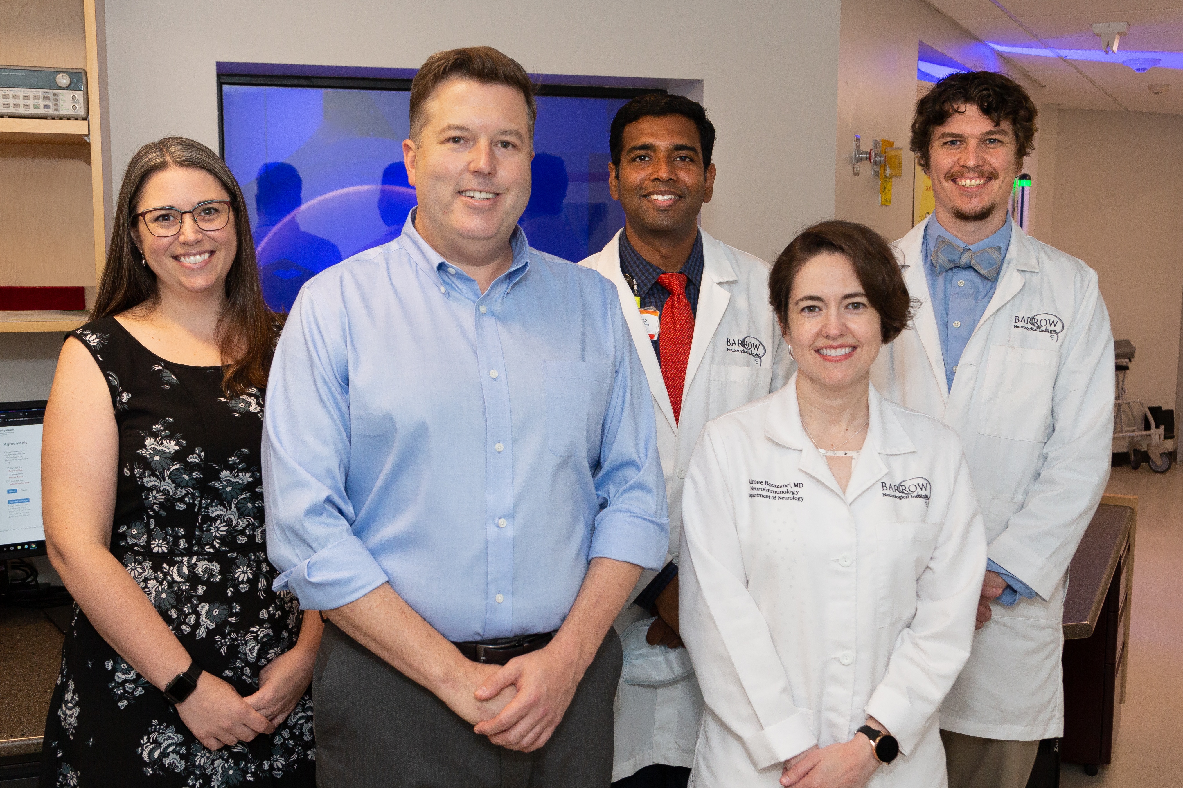
As part of his work in the MS field, Dr. Dortch partnered with Barrow imaging scientist Ashley Stokes, PhD, and MS neurologists Aimee Borazanci, MD, Ram Narayan, MD, and Michael Robers, MD, to apply to the North American Imaging in MS (NAIMS) Cooperative.
The organization, which serves as a network of academic centers across the United States and Canada, welcomed Barrow in June. The Institute is one of only two NAIMS member sites in Arizona.
“The focus is to share different emerging imaging tools and to try to standardize these methods for large-scale clinical trials,” Dr. Dortch said. “We’re excited to be a part of it. It helps raise our visibility in the field and allows us to participate in larger multi-site studies.”
A Point of Pride: Peripheral Nerve Imaging
Other research accomplishments in which Dr. Dortch takes great pride involve peripheral nerve regeneration imaging.
The cause-and-effect relationship between peripheral nerve injury and functional deficit appealed to Dr. Dortch as an engineer. He then discovered that he’d tapped into an understudied area with enormous potential to help improve patients’ lives.
“Peripheral nerves are just fascinating,” he said. “They can regrow, unlike central nerves.”
But because peripheral nerves regenerate slowly, about an inch per month, physicians have historically relied on a “wait-and-see” approach to gauge patient outcomes. It could take months or even years to determine if a patient has benefited from a nerve repair procedure.
Dr. Dortch aims to develop and validate new MRI-based biomarkers so that physicians can monitor nerve regeneration as it happens and re-operate before it’s too late to change the patient’s outcome.
Before arriving at Barrow, Dr. Dortch focused on injuries to the peripheral nerves themselves. Now, he’s also exploring whether he can apply his imaging methods to nerve transfers.
Recent advancements in nerve transfer surgery are enabling neurosurgeons, like Dr. Rory Murphy at Barrow, to restore hand function in patients who have sustained a spinal cord injury in the neck.
Nerve transfer involves taking a nerve from above the level of injury that is not critical to an area’s function, or that is redundant to another nerve, and attaching it to a significant nerve below the level of injury. The healthy donor nerve can then take over the function of the non-functioning peripheral nerve over time.
Not only could Dr. Dortch’s MRI techniques help detect failed nerve transfers in a timely manner, but they could help surgeons select the ideal donor nerve for each patient.
Measuring Success: Real-World Impact
Whether he is advancing neuroimaging through his own research, or mentoring trainees who then take their knowledge out into the world, Dr. Dortch says a successful career will be one where he is able to have a broad, real-world impact.
“We want to validate our methods here but then eventually push them out so that no matter where you go, you have access to these tools that can improve your care and ultimately quality of life,” he said.