Pineal Cyst
At a Glance
- A pineal cyst is a small, fluid-filled sac within the pineal gland, a small endocrine gland in the center of the brain.
- Most pineal cysts are benign and don’t cause symptoms; however, larger ones may lead to headaches, visual disturbances, balance issues, nausea, sleep disturbances, or cognitive changes.
- Doctors typically monitor pineal cysts with routine imaging and recommend surgical intervention only when symptoms are significant.
Overview
A pineal cyst is a small, fluid-filled sac that forms in the pineal gland, a tiny gland located deep in the brain. This gland helps regulate sleep by producing the hormone melatonin. Because pineal cysts often don’t cause symptoms, they are frequently discovered incidentally during brain imaging. In most cases, these cysts are harmless and don’t require treatment.
However, in rare instances, if a cyst grows large enough, it might lead to headaches or other neurological symptoms. If you’re diagnosed with a pineal cyst, your doctor may recommend regular check-ups to monitor its size, but treatment is not necessary for most people.
About the Pineal Gland
The pineal gland is a small, almond-shaped structure located deep inside the brain, between the left and right hemispheres. Roughly the size of a pea, the pineal gland helps regulate the body’s sleep-wake cycle by producing a hormone called melatonin. Melatonin is released in response to changes in light, signaling to the body when it’s time to sleep and when it’s time to be awake. This tiny gland plays a key role in maintaining your body’s natural rhythms, influencing not just sleep but also overall health and well-being.

What causes pineal cysts?
The exact cause of pineal cysts isn’t entirely clear, but they are generally thought to result from changes in the pineal gland’s structure or function. Some experts believe they may form during early development in the womb, where fluid-filled spaces naturally occur in the gland. Over time, these spaces might expand, leading to cyst formation.
In some cases, hormonal changes related to the pineal gland’s melatonin production could play a role. Age-related degeneration or natural wear and tear of the gland could also contribute to the formation of these cysts. Despite these theories, most pineal cysts are not linked to any specific external cause or underlying medical condition and often occur without any symptoms or health consequences.

Pineal Cyst Symptoms
The symptoms of a pineal cyst can vary depending on its size and whether it is pressing on nearby parts of the brain. Many people with a pineal cyst don’t experience any symptoms, especially if the cyst is small. However, some symptoms may occur if the cyst grows or affects the surrounding structures.
Symptoms of a pineal cyst can include:
- Headaches: Persistent headaches that feel like pressure in your head can signify a pineal cyst. Sometimes, they may worsen when lying down, as changes in brain fluid pressure can aggravate the discomfort.
- Vision Problems: Large pineal cysts can press on brain areas responsible for vision. You might notice blurred vision, double vision, or difficulty focusing. Some people also report an unusual sensitivity to bright lights.
- Dizziness and Balance Issues: If the cyst impacts brain areas involved in balance or coordination, you could feel dizzy, unsteady, or have trouble walking straight.
- Sleep Disturbances: A cyst might disrupt sleep patterns since the pineal gland helps regulate sleep through the hormone melatonin. You could experience difficulty falling asleep, waking up frequently, or feeling overly tired during the day.
- Nausea and Vomiting: If the cyst causes increased pressure in the brain, it might lead to nausea or vomiting. This is more common in cases where the cyst blocks the normal flow of cerebrospinal fluid, leading to hydrocephalus (increased fluid pressure in the brain).
- Memory or Focus Problems: Some people report concentration or short-term memory issues. This can happen if the cyst is causing pressure or disrupting communication between brain areas.
- Rare and Severe Symptoms: In rare cases, a pineal cyst can grow large enough to cause serious issues, including seizures, extreme difficulty with coordination, or a significant loss of consciousness. Such symptoms are medical emergencies.
If you’re experiencing any of these symptoms, it doesn’t necessarily mean you have a pineal cyst. Many of these symptoms can have other causes, so it’s essential to consult a doctor.
Diagnosis
Physicians typically diagnose pineal cysts using imaging tests, often when attempting to diagnose unrelated symptoms or performing a scan for another reason. The most common diagnostic tests for a pineal cyst include:
- Initial Evaluation: If you have frequent headaches, vision changes, or sleep disturbances, your doctor will likely start by asking about your medical history and symptoms. Based on this, they might suspect something involving the brain and recommend further testing.
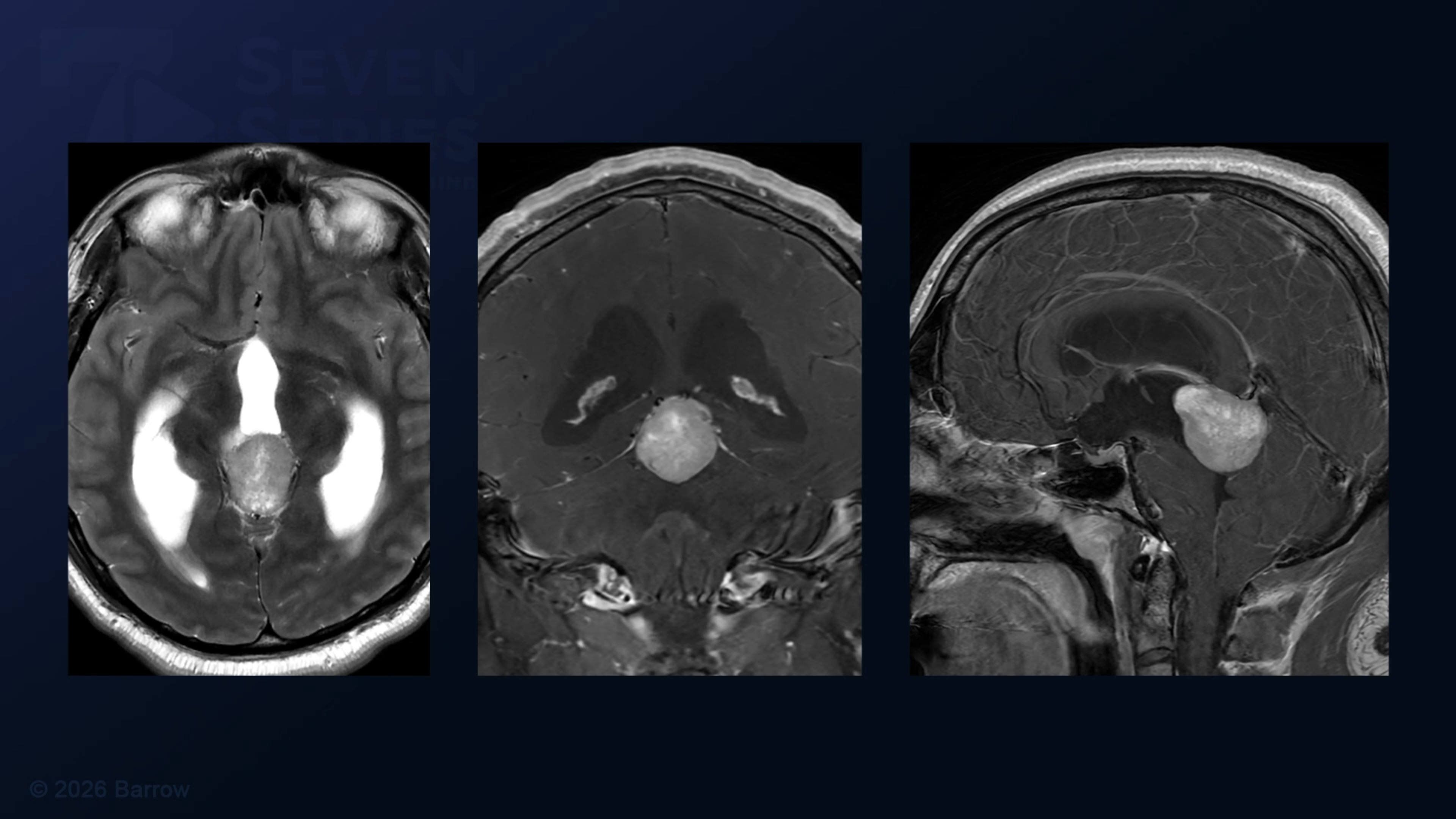
- Magnetic Resonance Imaging (MRI): The most common diagnostic test for a pineal cyst, an MRI uses strong magnets and radio waves to provide detailed brain and spinal cord images to determine a lesion’s location, size, and characteristics. Pineal cysts typically appear as well-defined, fluid-filled sacs on an MRI scan.
- Computed Tomography (CT) Scan: CT scans rely on X-rays to create detailed cross-sectional images of the brain, helping to detect cysts while evaluating their size and location. In some cases, a CT scan may show a pineal cyst, especially if there’s evidence of hydrocephalus in the brain.
- Monitoring and Follow-Up: Your care team may not recommend immediate treatment if a pineal cyst is not causing symptoms. Instead, they might monitor the cyst with follow-up MRI scans to ensure it isn’t growing or causing new symptoms. Depending on the size and appearance of the cyst, these follow-ups could happen every 6–12 months.
If a cyst is large or causing symptoms, your doctor might order more tests to evaluate its impact. For example:
- Eye exams to check for vision changes
- Neurological exams to assess balance, coordination, and reflexes
- Pressure monitoring if there are signs of increased pressure in the brain, like hydrocephalus
Diagnosing a pineal cyst is generally straightforward, but the decision to monitor or treat it depends on the size of the cyst, its growth, and any symptoms you might have.

Pineal Cyst Treatment
Treatment for a pineal cyst depends on whether it is causing symptoms. Small, asymptomatic cysts don’t normally require treatment. Doctors typically recommend monitoring it over time with regular brain scans to ensure it isn’t growing or causing problems.
Treatment might be necessary if the cyst is causing symptoms like headaches, vision problems, or increased pressure in the brain. A neurosurgeon can remove the cyst or create a way for fluid to drain, relieving pressure in the brain.
In rare cases where the cyst blocks the normal flow of brain fluid and causes hydrocephalus, doctors may place a small tube (called a shunt) to help drain the fluid and reduce pressure.
Treatment is always personalized. Your doctor will weigh the risks and benefits of different approaches and work with you to decide the best course of action based on your symptoms and overall health.
Nonsurgical Treatments
In most cases, nonsurgical treatments are unnecessary for pineal cysts. However, if the cyst is causing mild or manageable symptoms, nonsurgical options may help alleviate discomfort or improve quality of life. These approaches focus on managing the symptoms rather than treating the cyst itself, and can include:
- Headache Relief: Over-the-counter pain relievers like ibuprofen or acetaminophen can help manage headaches associated with a pineal cyst. If the headaches are severe or chronic, a doctor might prescribe stronger medications, such as triptans or other migraine-specific treatments.
- Sleep Support: Since the pineal gland regulates sleep through melatonin production, your doctor may address sleep disruption with melatonin supplements or other sleep aids. Relaxation techniques, improved sleep hygiene, and managing stress can also support better sleep.
- Vision Issues: If vision problems are mild, your eye doctor might recommend glasses, eye exercises, or other interventions to reduce strain.
- Hydrocephalus Symptoms: If there are early signs of increased pressure in the brain but surgery isn’t immediately necessary, a doctor might recommend diuretics or medications to reduce fluid buildup temporarily. However, this is not a long-term solution and requires close monitoring.
- Lifestyle Adjustments: Some people find relief by addressing contributing factors that may worsen symptoms, such as reducing stress, staying hydrated, maintaining a healthy diet, and avoiding excessive caffeine or alcohol intake.
Surgery
Neurosurgery for a pineal cyst is typically only recommended if the cyst is causing significant symptoms, such as severe headaches, vision disturbances, or increased pressure in the brain (hydrocephalus). The procedure is delicate because the pineal gland is located deep within the brain, near critical structures.
The most common procedure is a craniotomy, where a small opening is made in the skull to access the cyst under general anesthesia. Modern techniques often involve minimally invasive approaches, using specialized tools and microscopes for precision.
Once your neurosurgeon accesses the cyst, it is removed entirely or drained of its fluid contents. If the cyst has caused hydrocephalus, the surgeon may also create a pathway for cerebrospinal fluid (CSF) to flow or place a ventriculoperitoneal shunt. This small tube drains excess fluid to another part of the body.
After surgery, you’ll be monitored in the intensive care unit (ICU) for the first 24–48 hours to ensure there are no complications, such as bleeding or swelling in the brain. You may experience some fatigue, mild headaches, or temporary vision changes, but these usually improve over time.
Most people stay in the hospital for a few days, depending on their recovery progress. Neurocritical care specialists will conduct physical and neurological exams regularly to check for improvement in symptoms like headaches or vision problems. If you’re considering surgery, your doctor will discuss the potential benefits, risks, and what to expect during recovery to help you make an informed decision.
Video: Surgical Removal of Pineal Tumor
Warning: Contains surgical footage. Viewer Discretion is advised.
Common Questions
How common are pineal cysts?
Pineal cysts are relatively common, especially when detected during brain imaging studies for other reasons. They are usually incidental findings, meaning doctors detect them while testing for another condition, not because the cyst causes symptoms.
Studies suggest that pineal cysts are found in up to 10–20% of people during MRI scans of the brain.
They are more common in adults but can also occur in children and adolescents.
What size pineal cyst should be removed?
The decision to remove a pineal cyst is not based solely on its size but on its symptoms, growth, and impact on surrounding structures. However, larger cysts are more likely to cause problems and may prompt surgical intervention.
A neurosurgeon may recommend removal if the cyst causes significant symptoms such as persistent headaches, vision disturbances, balance issues, or hydrocephalus (increased pressure in the brain due to blocked cerebrospinal fluid flow). These symptoms often occur with cysts larger than 1–2 centimeters in diameter.
While smaller cysts (under 1 cm) are usually harmless, cysts measuring over 2 centimeters are more likely to be symptomatic and may warrant closer monitoring or removal.
Cysts larger than 3 centimeters are rare but often require surgery because they are at a higher risk of causing pressure-related symptoms.
A cyst that grows significantly over time, even if it starts small, may indicate a need for removal, especially if it begins causing symptoms.
Regardless of size, removal may be necessary if the cyst compresses nearby areas, such as the tectal plate (affecting vision or balance), or blocks the flow of cerebrospinal fluid.
Cysts with irregular or unusual imaging features may raise concerns about possible malignancy (such as a tumor). They might require surgical removal for biopsy and diagnosis.
Can pineal cysts become cancerous?
Pineal cysts are generally non-cancerous (benign) and rarely become cancerous. In most cases, they remain stable and do not pose a significant risk. However, it’s essential to distinguish pineal cysts from other growths in the pineal gland, as true tumors of the pineal region (such as pineocytomas, pineoblastomas, or germ cell tumors) can occur, although they are much rarer.
What is the prognosis for a pineal cyst?
The prognosis for a pineal cyst is generally excellent, especially in cases where the cyst is small, asymptomatic, and stable. Most people with pineal cysts live normal, healthy lives without any complications or need for treatment. The prognosis is usually very good for patients who undergo surgery to remove a symptomatic pineal cyst. Modern surgical techniques are highly effective, and most people experience significant relief from symptoms after the cyst is removed. Recovery from surgery typically takes a few weeks, and long-term complications are rare when performed by an experienced neurosurgeon.