
Clinical Images: A Self-Inflicted Crossbow Wound
Authors
Randall W. Porter, MD
Carlos A. Carrion, MD
Dean G. Karahalios, MD
Division of Neurological Surgery, Barrow Neurological Institute, Mercy Healthcare Arizona, Phoenix, Arizona
Key Words : crossbow wound, frontotemporal
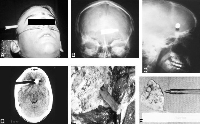
A 16-year-old Caucasian male sustained a self-inflicted crossbow wound to the right frontotemporal region (Panel A). He was intact on neurological examination and had no significant medical history. Plain anteroposterior (Panel B) and lateral (Panel C) radiographs of the skull confirmed that the arrow had entered the right frontal region and crossed the midline into the left frontal region. The long end of the arrow was transected approximately 5 cm from its entrance into the skull so that the patient’s head could fit in the computed tomography scanner. Computed tomography of the brain showed no significant hemorrhage but confirmed the trajectory of the arrow from the right frontal to the left frontal region (Panel D). Angiography revealed no significant vessel occlusion or laceration.
The patient was taken to the operating room where a bicoronal craniotomy was performed to achieve proximal and distal exposure. This procedure was followed by a right frontotemporal craniotomy around the arrow (Panel E). The arrow and bone flap were removed together. Hemostasis was achieved, the dura closed, and the bone flap replaced. The arrow was sent to pathology for examination(Panel F). The patient was neurologically intact after the operation and made an uneventful recovery.