Spinal Disk Arthroplasty
Authors
Mark S. Gerber, MD
Robert M. Galler, DO
Stephen M. Papadopoulos, MD
Division of Neurological Surgery, Barrow Neurological Institute, St. Joseph’s Hospital and Medical Center, Phoenix, Arizona
Abstract
The cervical and lumbar portions of the spine are frequently fused to treat instability and degenerative disease of the spine. As a result of alterations in the natural biomechanics of the spine, such patients have a high risk of developing progressive spondylotic disease at levels adjacent to the levels fused. To decrease the risk of further degeneration, ongoing research has sought to create a functional prosthetic disk that preserves motion at the diseased level thereby resolving adjacent level arthrosis physiologically. This article reviews the history and development of the artificial disk and its status in human trials.
Key Words: artificial disk, adjacent segment disease, spinal arthroplasty
During the last 50 years, the treatment of symptomatic degenerative spinal disease has evolved significantly. Despite advances in techniques for cervical and lumbar surgery, opinions about the overall long-term success of these operations continue to differ. Hilibrand et al.[12] found that 19% of patients had adjacent segment disease 10 years after undergoing anterior cervical fusion. Hilibrand et al.[12,13] reported a 3% per year rate of symptomatic disk degeneration at levels adjacent to cervical fusion. Others have found that 7 to 15% of patients need a reoperation at adjacent levels.[1,5,10,15] Considering the overall excellent fusion rates and outcomes associated with anterior cervical fusion, adjacent segment disease is likely to be an ongoing problem for patients and surgeons.
Lumbar fusion effectively relieves pain by eliminating motion due to segmental instability, by restoring disk and foraminal height, and by halting the progression of degeneration at the treated level. However, evidence suggests that these procedures also may accelerate the development of adjacent segment disease. In one series, 50 to 60% of the patients had persistent back pain and 20 to 30% had recurrent radiculopathy 10 years after undergoing lumbar diskectomy.
From a biomechanical perspective, adjacent level disease is a relatively easy concept to understand. After the cervical or lumbar vertebra fuse, the fused vertebral bodies create a longer lever arm. The load forces once shared across the fused bodies now concentrate on either end of the fusion mass, thereby accelerating the degeneration of these joints. DiAngelo et al.[6] have shown that fusion increases local motion in adjacent segments as well as global cervical motion.
The idea of joint replacement for the treatment of degenerative disease revolutionized the practice of orthopedic surgery. It is possible that disk arthroplasty may similarly affect the treatment of spondylotic disease of the spine. This initiative has motivated a significant portion of the impetus to develop artificial disks.
History of Disk Replacement
In 1966 Fernstrom[8] published his experience with 199 intracorporal endoprostheses implanted into 125 patients and followed for 4 to 7 years. The prosthesis was a corrosion-resistant stainless steel ball placed in the center of an evacuated disk space. Prosthetics of different sizes were implanted in both the cervical and lumbar spine. An anterior approach was used for the cervical implantations, and a laminectomy was used to access the lumbar spine. During the follow-up period, subsidence occurred in 88% of the patients leading to the abandonment of the technique.
At the First International Symposium on the Artificial Disk held in Berlin in 1989, only a few artificial nucleus designs were presented. Since then research and development of artificial disks have blossomed. Overseas, the Conformité Europeene has already approved several replacement disks. Consequently, most of the clinical data and experience come from these centers experimenting and developing this technology.
Prosthetic Design
The functions of the disk are to prevent collapse of the vertebral bodies, to maintain motion, to insure stability, and to reduce pain. Using technology gained from orthopedic joint replacements, numerous strategies have been devised to create a successful prosthetic disk. Elastomers, viscous fluids, fluid-filled chambers, and articulating components have all been entertained. Strength, durability, and biomechanical and biochemical compatibility must be considered. Furthermore, ease of implantation and the immediate postoperative and long-term stability of the device will affect the utility of a replacement disk. Wear can cause debris to accumulate within the artificial joint space, causing the joint to fail prematurely and disrupting the interface between materials such as metal-polymer, metal-metal, and metal-bone. Joint stability also is greatly affected by the ability of the joint to fuse with adjacent bony surfaces. Based on this information, prosthetics disks can be divided into two major categories: nucleus replacements and total disk replacements.

Nucleus Replacement
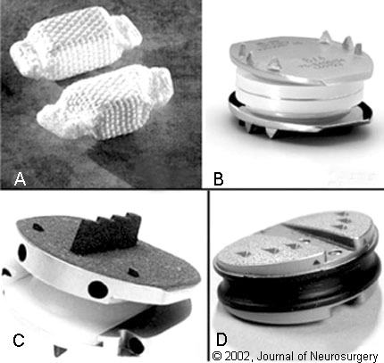
Nucleus replacements are designed for use when the major feature of the degenerative process involves the nucleus pulposus but has spared the annulus and supporting structures. The Prosthetic Disc Nucleus (PDN, Raymedica, Bloomington, MN) is an implant with a hydrogel core wrapped in a woven polyethylene cover (Fig. 1). A dehydrated spacer, it expands as it absorbs water after implantation. The size of the implant varies. It can be placed through an anterior or posterior approach. For proper balance it should be used in pairs. Outcomes of a cadaveric study of the biomechanics of this implant were favorable. The cadaveric model, however, failed to permit full hydration of the device.19 Since 1996, 423 patients have received the PDN since 1996 with a surgical success rate of 90%. Clinical results have been encouraging. The main problem, the 10% migration rate, has led to a series of modifications to the procedure.[14]
Outcomes are not yet available for two other nucleus replacements. Aquarelle Stryker (Howmedica, Rutherford, NJ) is a hydrogel material based on polyvinyl alcohol, which is hydrated before implantation. This material can be implanted through a tapered cannula placed in an annulotomy via a posterior or lateral approach. The Prosthetic Intervertebral Nucleus (Raymedica, Bloomington, MN) is a polyurethane material instilled into a balloon that is inserted into a disk space. Once placed into the disk space, the chamber is filled with the material, which then cures in situ.
Total Disk Replacement
In contrast to nucleus replacement, total disk replacement addresses degenerative processes throughout the disk and associated structures. It represents a major reconstructive procedure. These prosthetics are designed to restore the normal movement of the diseased motion segment. In the lumbar spine, the Link SB Charité III (Waldemar Link GmbH and Co., Hamburg, Germany), Acroflex-100 prosthesis (DePuy AcroMed, Raynham, MA), and Prodisc (Aesculap AG and Co., Tuttlingen, Germany) are the most widely implanted total-disk replacements (Fig. 1). The Cummins/Bristol disk (Medtronic Sofamor Danek, Memphis, TN) and the Bryan Cervical Disc Prosthesis (Medtronic Sofamor Danek, Memphis, TN) are under investigation for use in the cervical spine.

Cervical Arthroplasty
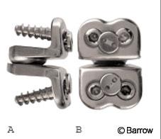
The Cummins/Bristol disk is a two-piece ball-and-socket device that is secured to the anterior vertebral bodies with screws (Fig. 2). Clinical outcomes were good in 26 patients followed for 2.4 years. Unfortunately, the stainless steel devices, which were manufactured at a local foundry, may have suffered from a production error. There were five cases of hardware failure, one of which required surgical revision. Wigfield et al.[17,18] have shown that the implantation of an artificial disk seems to decrease adjacent level motion compared with fusion.


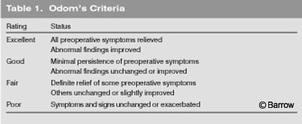
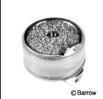
In Europe the Bryan Cervical Disc (Medtronic Sofamor Danek, Memphis, TN; Fig. 3) prosthesis has been implanted in 97 patients, and results have been promising. At the time of writing, 30 patients have been followed for 1 year, and 67 patients have been followed for 6 months. The clinical success rate was 90% and 86%, respectively, based on Odom’s criteria of excellent, good, or fair (Table 1). Long-term follow up is needed to determine whether the disks remain functional and what effect they have on adjacent levels.[9]
Lumbar Arthroplasty
In 1984 Drs. Karin Büttner-Janz and Kurt Schellnack began to work with the Waldemar Link GmbH & Company (Hamburg, Germany) to develop a replacement lumbar disk. After several design iterations, the final version of the disk, the LINK SB Charité III, is currently available in Europe.[2,3,11] The device is composed of a ultrahigh molecular weight core sandwiched between two cobalt-chromium alloy endplates. More than 3000 of these disks (all generations) have been implanted throughout Europe.[20] In 1994 a multicenter retrospective review of the early clinical results with the LINK SB Charité lumbar artificial disk was published.[11] Compared to their preoperative condition, pain, mobility, walking distance, and weakness improved significantly in 93 patients. The complication rate, as a result of disk migration or dislocation and device failure, was 6.5%. Cinotti et al.[4] and Zeegers et al.[21] also reported their experiences with the LINK SB Charité III disk. Forty-six patients were studied a mean of 3.2 years after implantation: 63% reported satisfactory results. The success rate was 69% in patients who underwent isolated disk replacement and 77% in patients who had undergone previous back surgery. Two patients had the prosthesis removed. Seven patients underwent posterolateral fusion without removal of the device.
Enker et al.[7] reported their experience with the Acroflex artificial lumbar disk. This prosthesis consists of two titanium endplates vulcanized to a polyolefin rubber core (Fig. 1). The disk was implanted in six patients via a midline transabdominal approach, four of whom reported satisfactory results. In one patient, the rubber core fractured and a revision was necessary.
The ProDisk, developed by Dr. Thierry Marnay, consists of two CrCoMo alloy endplates covered with titanium plasmopore to enhance osteointegration (Fig. 1). The inferior articulation is a monoconvex surface that slides into a concave upper rim. This design permits the implant to be inserted with significantly less distraction of the disk space than the LINK SB Charité III disk. Because movement is across two surfaces, the motion is semiconstrained. The clinical outcomes associated with the ProDisk have been presented at several meetings, but published data are limited.[16]
Risks of Artificial Disk Replacement
A major concern of artificial disk replacement is that of implant migration. In the cervical spine, disk migration can have grave consequences whereas more leeway exists in the lumbar spine. Because most neurosurgeons are familiar with the anterior approach to the cervical spine, the first prosthesis to be approved by the Food and Drug Administration (FDA) will likely be for use in the cervical spine. Furthermore, cervical spondylosis is often attacked through an anterior approach. It should be possible to implant an artificial disk in the lumbar spine through an anterior or retroperitoneal approach. A posterior approach, performed similar to a posterior lumbar interbody fusion, also may be possible. From a technical perspective, advancing and anchoring a disk from a posterior approach will be difficult if the artificial disk replacements for the lumbar spine remain in their current configuration.
Longevity and choice of materials are other issues that need to be addressed. Most of the present artificial disk replacements have been modified in some fashion. In fact, some artificial disk replacements have failed. Automated test jigs permit relatively straightforward testing of artificial disk replacements for fatigue and other biomechanical features.
Status in the United States
The hurdles to FDA approval for these devices are numerous. Obvious challenges include the use of biocompatible materials. Given the present data on many metals and other composite materials already used in joint prosthesis, this bar should not be too difficult to overcome. Other issues include the perceived patient risk-to-benefit ratio. In particular, artificial disk replacement must be shown to be safe and effective, especially when many alternatives for the treatment of degenerative disk disease already exist. Furthermore, the environment in the United States, highlighted by the litigations over pedicle screws and breast augmentation, mandates that these devices be tested rigorously before they are marketed. Of great concern is the risk of disk migration and the potential for devastating neurologic injury.
At present in the United States several artificial disks are undergoing testing in humans. The LINK SB Charite III disk and the Prodisc have already been implanted at several centers. The Investigational Device Exemption Study for the Charite III has recently been completed and is still in progress for the ProDisk. The Bryan Cervical Prosthetic Disk is to begin testing in the United States in the near future.
Discussion
Anterior cervical diskectomies are among the most common spinal procedure performed by neurosurgeons. Each year more than 35,000 Americans undergo diskectomy and fusion. Most patients do very well after this procedure. Complication rates are low, and the associated symptoms tend to improve significantly. Lumbar fusion also is used widely, but its rate of clinical success is more variable.
Considering that most neurosurgeons are familiar and comfortable with the anterior cervical approach, anterocervical disk replacement is likely to be the first location that will enjoy the benefit of this new technique. In contrast, most lumbar procedures are performed with the patient in a prone position. Placing an artificial disk into the lumbar spine from this position would be exceedingly difficult and would likely require a lateral approach. Otherwise, retroperitoneal or anterior approaches, which tend to be associated with relatively high rates of morbidity and mortality, will need to be used.
Given the increased interest in minimally invasive spinal surgery, it will be interesting to see how these two strategies compete with one another. Surgeons know that the paraspinous muscles confer a significant amount of strength and stability and that stripping these muscles from the spinous processes increases the loads on the anterior longitudinal ligament and facet joints. Minimally invasive techniques can be combined with spinal arthroplasty to minimize procedure-related morbidity while maximally preserving the functional integrity of the spine.
The treatment of spinal disease with arthroplasty continues to evolve in a way that recalls the different methods of joint replacement developed in orthopedic surgery. The optimal treatment of spinal spondylosis will likely be reconstituting the physiological state of motion rather than fusing and immobilizing the joints involved.
References
- Baba H, Furusawa N, Imura S, et al: Late radiographic findings after anterior cervical fusion for spondylotic myeloradiculopathy. Spine 18:2167- 2173, 1993
- Büttner-Janz K, Schellnack K: Principle and initial results with the Charité Modular type SB cartilage disk endoprosthesis [Hungarian]. Maagyar Traumatologia, Orthopaedia Es Helyreallito Sebeszet 31:136-140, 1988
- Büttner-Janz K, Schellnack K, Zippel H: An alternative treatment strategy in lumbar intervertebral disk damage using an SB Charité modular type intervertebral disk endoprosthesis [German]. Zeitschrift fur Orthopadie und Ihre Grenzgebiete 125:1-6, 1987
- Cinotti G, David T, Postacchini F: Results of disc prosthesis after a minimum follow-up period of 2 years. Spine 21:995-1000, 1996
- Clements DH, O’Leary PF: Anterior cervical discectomy and fusion. Spine 15:1023-1025, 1990
- DiAngelo DJ, Foley KT, Vossel KA, et al: Anterior cervical plating reverses load transfer through multilevel strut-grafts. Spine 25:783-795, 2000
- Enker P, Steffee A, Mcmillin C, et al: Artificial disc replacement. Preliminary report with a 3-year minimum follow-up. Spine 18:1061-1070, 1993
- Fernstrom U: Arthroplasty with intercorporal endoprothesis in herniated disc and in painful disc. Acta Chir Scand Suppl 357:154-159, 1966
- Goffin J, Casey A, Kehr P, et al: Preliminary clinical experience with the Bryan cervical disc prosthesis. Neurosurgery 51:840-817, 2002
- Gore DR, Sepic SB: Anterior cervical fusion for degenerated or protruded discs. A review of one hundred forty-six patients. Spine 9:667-671, 1984
- Griffith SL, Shelokov AP, Büttner-Janz K, et al: A multicenter retrospective study of the clinical results of the LINK® SB Charite intervertebral prosthesis. The initial European experience. Spine 19:1842-1849, 1994
- Hilibrand AS, Carlson GD, Palumbo MA, et al: Radiculopathy and myelopathy at segments adjacent to the site of a previous anterior cervical arthrodesis. J Bone Joint Surg Am 81:519-528, 1999
- Hilibrand AS, Yoo JU, Carlson GD, et al: The success of anterior cervical arthrodesis adjacent to a previous fusion. Spine 22:1574-1579, 1997
- Klara PM, Ray CD: Artificial nucleus replacement. Clinical experience. Spine 27:1374-1377, 2002
- Lunsford LD, Bissonette DJ, Jannetta PJ, et al: Anterior surgery for cervical disc disease. Part 1: Treatment of lateral cervical disc herniation in 253 cases. J Neurosurg 53:1-11, 1980
- Traynelis V: Spinal arthroplasty. Neurosurg Focus 13:Article 10, 2002
- Wigfield C, Gill S, Nelson R, et al: Influence of an artificial cervical joint compared with fusion on adjacent-level motion in the treatment of degenerative cervical disc disease. J Neurosurg 96:17-21, 2002
- Wigfield CC, Robertson J, Metcalf N, et al: The influence of an artificial cervical joint versus fusion on adjacent level motion in the treatment of cervical disc disease (abstract). Neurosurgery 47:516, 2000
- Wilke HJ, Kavanagh S, Neller S, et al: Effect of a prosthetic disc nucleus on the mobility and disc height of the L4-5 intervertebral postnucleotomy. J Neurosurg 95:208-214, 2001
- Yuan HA, Bao Q-B: Disc arthroplasty. SpineLine November/December:6-11, 2001
- Zeegers WS, Bohnen LM, Laaper M, et al: Artificial disc replacement with the modular type SB Charité III: 2-year results in 50 prospectively studied patients. Eur Spine J 8:210-217, 1999
