
How Are Feelings Represented in the Human Brain?*
A. D. (Bud) Craig, PhD
Division of Neurobiology, Barrow Neurological Institute, St. Joseph’s Hospital and Medical Center, Phoenix, Arizona
*Courtesy of A. D. (Bud) Craig, PhD
The Atkinson Research Laboratory
A. D. (Bud) Craig received his PhD in neurobiology from Cornell University in 1978. He studied at Washington University School of Medicine and at the University of Wurzburg (Germany) before arriving at Barrow in 1986. Others in this laboratory include the following: John Beggs obtained his PhD from Arizona State University in 1974 and has performed electron microscopic analyses at Barrow since then. Irina Strigo completed her PhD at McGill University in 2002, where she studied the functional imaging of pain in the laboratory of Catherine Bushnell. Karl Krout obtained his PhD at Washington University of School Medicine in 2001, where he studied the neuroanatomy of homeostatic systems with Arthur Loewy. Irina and Karl joined our laboratory in 2003. Le Hua completed her BS in biology at the University of Arizona in 2003 and performed fMRI studies with us before starting her medical training at the University of Southern California in 2004-2005. Leanne Brady received her BS in biology at the University of Notre Dame in 2001. Misty Auldridge received her BS in chemistry from Northern Arizona University in 2002. Leanne and Misty serve as technicians, and both began part-time training to become registered nurses in April 2005.
We use anatomical, physiological, and imaging methods to study the ways the brain represents the feelings we feel from our bodies, how these areas are interconnected, and how these areas are involved in emotions and mood disorders.
Key Words: depression, homeostasis, pain, temperature
Abbreviations used: fMRI, functional magnetic resonance imaging; PET, positron emission tomography; VMpo, posterior ventromedial nucleus
As humans, we perceive feelings from our bodies that relate our state of well-being, our energy and stress levels, our mood and disposition. This laboratory studies the functional anatomy of the brain’s representations of feelings from our bodies.
The most basic feelings from our bodies include sensations from the skin (like touch, temperature, pinch), feelings from our gut (like hunger, colon distension), feelings from our muscles and joints (ache, burn), and many other distinct feelings (toothache, thirst, vasomotor flush, sensual contact). The feelings from our bodies are directly related to our health, and so we tell each other every day “how we feel.”
The basic feelings from our bodies seem to be closely related to all of our other “feelings,” that is, our emotions (e.g., anger, sadness, happiness, fear, disgust). More than 100 years ago it was proposed that our bodily feelings provide the basis not only for emotions but also for our awareness of ourselves as “sentient” (or, feeling) beings. In fact, recent theoretical considerations and evidence from patients with brain damage argue that the portions of the brain that generate such feelings also engender subjective consciousness. The work in our laboratory is relevant to such large issues. However, it also has more immediate clinical significance for our understanding of the neural mechanisms underlying chronic pain, intractable itch, psychosomatic disorders, and major depression.
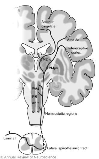
For almost 20 years, our work has focused on dissecting the specific connections from the spinal cord to the brain that carry activity important for bodily feelings in experimental animals. (Fig. 1). We perform several different types of anatomical and physiological experiments in animals. Increasingly, our recent work relies on psychophysical and functional imaging studies to analyze the activation pathways in humans.
Anatomical Studies
At present we are conducting several anatomical studies: (1) analysis of the connections of the region in the thalamus called VMpo with the cortex, (2) retrograde analysis of the neurons in the spinal cord that connect with the entire thalamus, and (3) analysis of the connections of the regions in the thalamus activated by stimulation of the vagus nerve.
In the first project, tracer chemicals are injected in the thalamus at the sites where neurons in anesthetized animals respond to thermal (cool, warm) or painful (hot, cold, pinch) stimulation. In this manner the cortical areas that receive such connections can be mapped. These areas include the “interoceptive cortex” and “area 3a” (Fig 1). Later, functional imaging in humans is used to verify the pathways identified by these anatomical experiments (see below).
Our prior work traced the ascending connections from the spinal cord to the thalamus and identified the region that we call VMpo as a critical site for the representation of temperature, pain, itch, and muscle feelings in primates and humans. The current project maps the next step in the progression of activity. It may reveal new features of the internal organization of the areas that represent these feelings in our brains.
In the second project, tracer chemicals are injected at various regions in the thalamus. The spinal cord neurons labeled with the tracer are mapped and thereby identified as having connections with the regions that contained injected tracer. This anatomical mapping procedure demonstrates the organization of the spinal connections with the thalamus. Thus, cells that project to parts of the thalamus involved in feelings of pain are directly associated with the generation of pain. Likewise, we infer that spinal cells that project to parts of the thalamus known to be involved in controlling movement have a role in motor control.
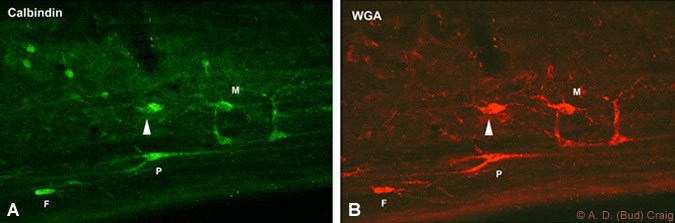
Although much is known about the properties of spinal cord neurons, these experiments are producing some surprising results. For example, many investigators studying pain believe that spinal cells in a region called lamina 5 are involved in pain. Our findings, however, indicate that these cells are much more likely to be involved with motoric reflexes because they overwhelmingly project to a region involved in motor control. In contrast, neurons in an area of the spinal cord called lamina 1 seem to be responsible for pain, temperature, itch, and other feelings from the body (Fig. 2). The reason for this discrepancy is that other investigators have confused the reflexive withdrawal initiated by a painful stimulus with the actual feeling of pain. Our work, however, indicates that the central nervous system distinguishes the motoric effects associated with pain from the actual feeling.
In the third project, tracers are injected in the thalamus at sites where activation by stimulation of the vagus nerve can be recorded. Vagal nerve stimulation is a new and simple therapeutic method that relieves treatment-resistant epilepsy and major depression. Nevertheless, the mechanisms underlying its efficacy are unknown. Our physiological (see below) and anatomical tracer studies are providing the fundamental knowledge needed to understand and to improve the efficacy of this treatment.
Physiological Studies
Several physiological projects complement the anatomical projects: (1) mapping the regions of the thalamus activated by vagal nerve stimulation, (2) recording the activity of single neurons in VMpo in response to various thermal and mechanical stimuli, and (3) recording the activity of single spinal cord neurons that send connections to different parts of the thalamus.
In the first project, the vagus nerve is stimulated in the same manner performed clinically, and a microelectrode is used to record evoked potentials in a three-dimensional grid across the thalamus. The sites activated by such stimulation can then be identified. This project is important because the literature indicates that in humans the thalamus is the key site activated by stimulation of the vagus nerve. The thalamus is an aggregate that contains many different regions that have very different connections with other parts of the brain. Therefore, far greater detail about where this vagal afferent activation occurs is needed to understand the clinical efficacy of stimulation of the vagus nerve. Our recent work has identified two activation sites. One of these sites may be important for the antiepileptic effects of vagal nerve stimulation, while the other may be important for the anti-depressive effects.
In the second project, microelectrode recordings from VMpo neurons in anesthetized animals are made and their activity is directly compared with the patterns of human feelings in response to the same stimuli. Similarly, in the third project, microelectrode recordings from spinal neurons are used to make similar comparisons. The important advantage is that physiological methods can also be used to directly identify the connections of the individual neurons from which we record. Thus, data can be collected to confirm or deny the hypothesized roles of these neurons in feelings from the body that are inferred from the results in the parallel anatomical experiments. Furthermore, the effects of various agents that modify (i.e., reduce) the activity of these neurons can be analyzed quantitatively.
Functional Imaging Studies
Several functional imaging investigations of humans are underway in our laboratory. These studies rely on fMRI, which allows observation of changes in blood flow within parts of the brain selectively activated by particular procedures. These experiments complement the anatomical and physiological studies in experimental animals. Four primary avenues of investigation are being conducted. First, regions associated with different feelings from the body in awake humans are identified and analyzed. Second, regions activated by thermal or painful stimuli in anesthetized monkeys are identified and analyzed. Third, patterns of activation by pain in normal humans are compared with the activation in depressed patients. Finally, the relationships between patterns of activation in the human brain under different conditions of emotion and cardiorespiratory state are examined.
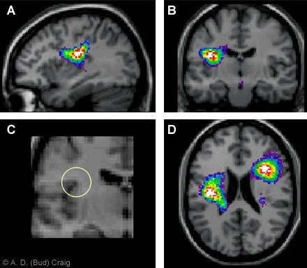
The first project builds on our prior anatomical work in monkeys and on the results of functional imaging in humans. These studies identified the main regions in the cortex involved in feelings from the body. In our earlier study, PET was used to identify areas of activation that correlated directly with graded cooling stimulation of the hand (Fig. 3). In our most recent observation, fMRI showed that this site in the brain precisely encodes the temperature of the stimulus applied to the body and also its location (Fig. 4). The spatial arrangement of these activation sites agrees with our anatomical and physiological studies.
Contrary to the prediction of earlier investigators, this location in the brain is not associated with touch or with control of movement. Rather, this site is located in a region associated with cardiorespiratory control. This finding underscores the evolutionarily primal importance of temperature (and other feelings from the body) for homeostasis, the dynamic process that maintains the integrity of the body. A stroke that damages this location in humans can cause an unremitting feeling of burning pain that even morphine cannot reduce. Our work provides a concrete explanation for this so-called “central pain” phenomenon as a homeostatic (thermoregulatory) dysfunction and suggests several possible new therapies.
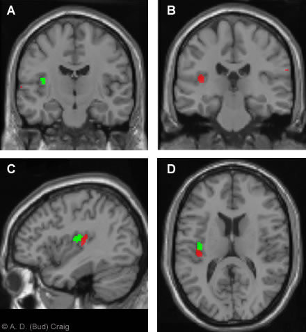
cool stimulus was applied to the hand (red) and neck (green).)
In the second project, fMRI studies of anesthetized monkeys directly complement the fMRI protocols in awake humans as well as the experimental physiological and anatomical studies in monkeys. This link is critical to validate the cross-primate comparisons of the activation of the brains of monkeys and humans.
The third and fourth projects are collaborative studies with colleagues at the University California, San Diego Medical School (led by Professor Martin Paulus, Department of Psychiatry) and at Arizona State University (led by Professor Alex Zautra, Department of Psychology). In the third project, fMRI is being used to examine the overlap and interdependence of brain regions activated by pain and by depression under conditions of cognitive distraction that constitute an emotional challenge for depressed patients. The fourth project is examining the hypothesis that the human forebrain has an asymmetric representation of emotions that is directly related to homeostatic function. This hypothesis extends our prior work on the subjective awareness of feelings from the body by contrasting positive and negative feeling states under different cardiorespiratory loads. An important potential implication of this work is the development of a biomarker for depression, which may be revealed by an imbalance of particular sites between the left and right sides of the forebrain.
Summary
These anatomical, physiological, and functional imaging projects complement each other and form a unified strategy to expand our knowledge about the representation of feelings from the body in the human brain. These studies will provide detailed insights into the organization of such regions and will extend our understanding of their participation in subjective emotional processes. The ability to make a cross-primate comparison provides concrete, precise anatomical descriptions of the regions activated in the human brain during these feelings. The ability to examine the activity in these regions in patients with emotional disorders provides insights into the interactions of feelings from the body with emotional state and could have direct clinical implications. We look forward to making further contributions that may help alleviate human pain and suffering.
Related Readings
- Craig AD: How do you feel? Interoception: the sense of the physiological condition of the body. Nat Rev Neurosci. 3:655-666, 2002
- Craig AD: Human feelings: why are some more aware than others? Trends Cognit Sci 8:239-241, 2004
- Craig AD: Distribution of trigemino- and spino-thalamic lamina I terminations in the macaque monkey. J Comp Neurol 477:119-148, 2004
- Hua LH, Strigo IA, Baxter LC, Johnson SC, Craig AD Craig: Antero-posterior somatotopy of innocuous cooling activation focus in human dorsal posterior insular cortex. Amer J Physiol Regul Integr Comp Physiol 289:R319, 2005