
Clinical Images: Persistent Primitive Trigeminal Artery with and without Aneurysm
Paul J. Apostolides, MD
Michael T. Lawton, MD†
Carlos A. David, MD
Robert F. Spetzler, MD
Division of Neurological Surgery, Barrow Neurological Institute, Mercy Healthcare Arizona, Phoenix, Arizona
†Current Address: Department of Neurosurgery, University of California-San Francisco, San Francisco, California
Key Words : trigeminal artery, anyeurysm
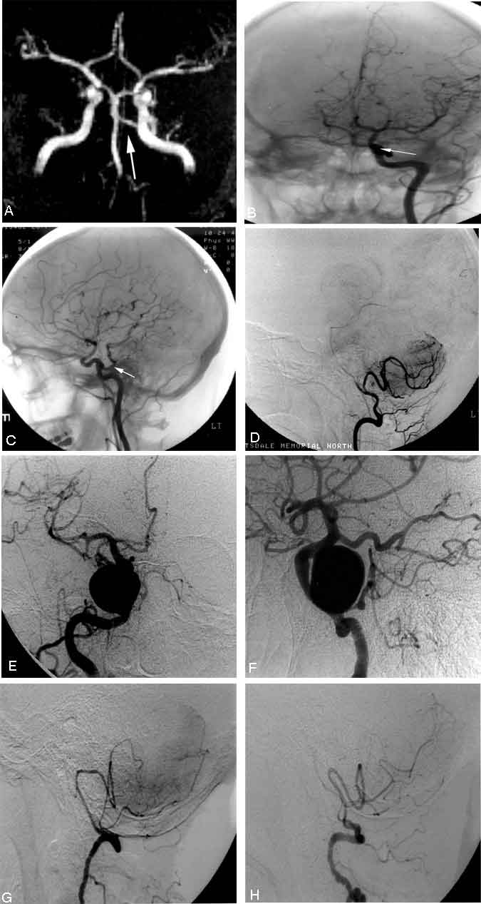
The primitive trigeminal artery, the most common of the persistent carotid-basilar anastomoses, is seen in about 0.6% of cerebral angiograms. Panel A demonstrates a primitive trigeminal artery ( arrow ) as seen on magnetic resonance angiography. The same primitive trigeminal artery ( arrow ) is visible on anteroposterior (AP, Panel B ) and lateral ( Panel C ) left common carotid artery angiograms. In this case, the posterior communicating arteries were hypoplastic, and the primitive trigeminal artery represented the main blood supply to the distal basilar artery, posterior cerebral artery, and superior cerebellar artery territories (Saltzman type I anatomy). The hypoplastic basilar artery is seen on a lateral view of a left vertebral artery angiogram ( Panel D ).
A primitive trigeminal artery is often associated with other cerebrovascular anomalies such as aneurysms and arteriovenous malformations, but aneurysms directly involving the primitive trigeminal artery, as seen on AP ( Panel E ) and lateral ( Panel F ) right common carotid artery angiograms of this case, are rare. In this patient, the posterior communicating arteries represented the main blood supply to the posterior cerebral arteries (Saltzman type II anatomy). As shown in lateral views of left ( Panel G ) and right ( Panel H ) vertebral artery angiograms, both vertebral arteries terminated as posterior inferior cerebellar arteries.