DoD Funds Barrow Neuroimaging Study of Peripheral Nerve Injury, Repair
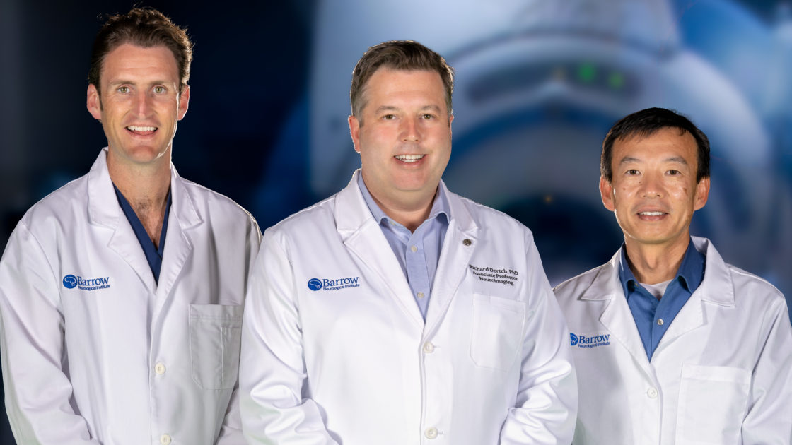
Richard Dortch, PhD, a neuroimaging researcher at Barrow Neurological Institute, has received a grant from the United States Department of Defense (DoD) to develop an imaging method to better diagnose peripheral nerve injuries and monitor recovery. This could help guide surgical decision-making, potentially improving patient outcomes.
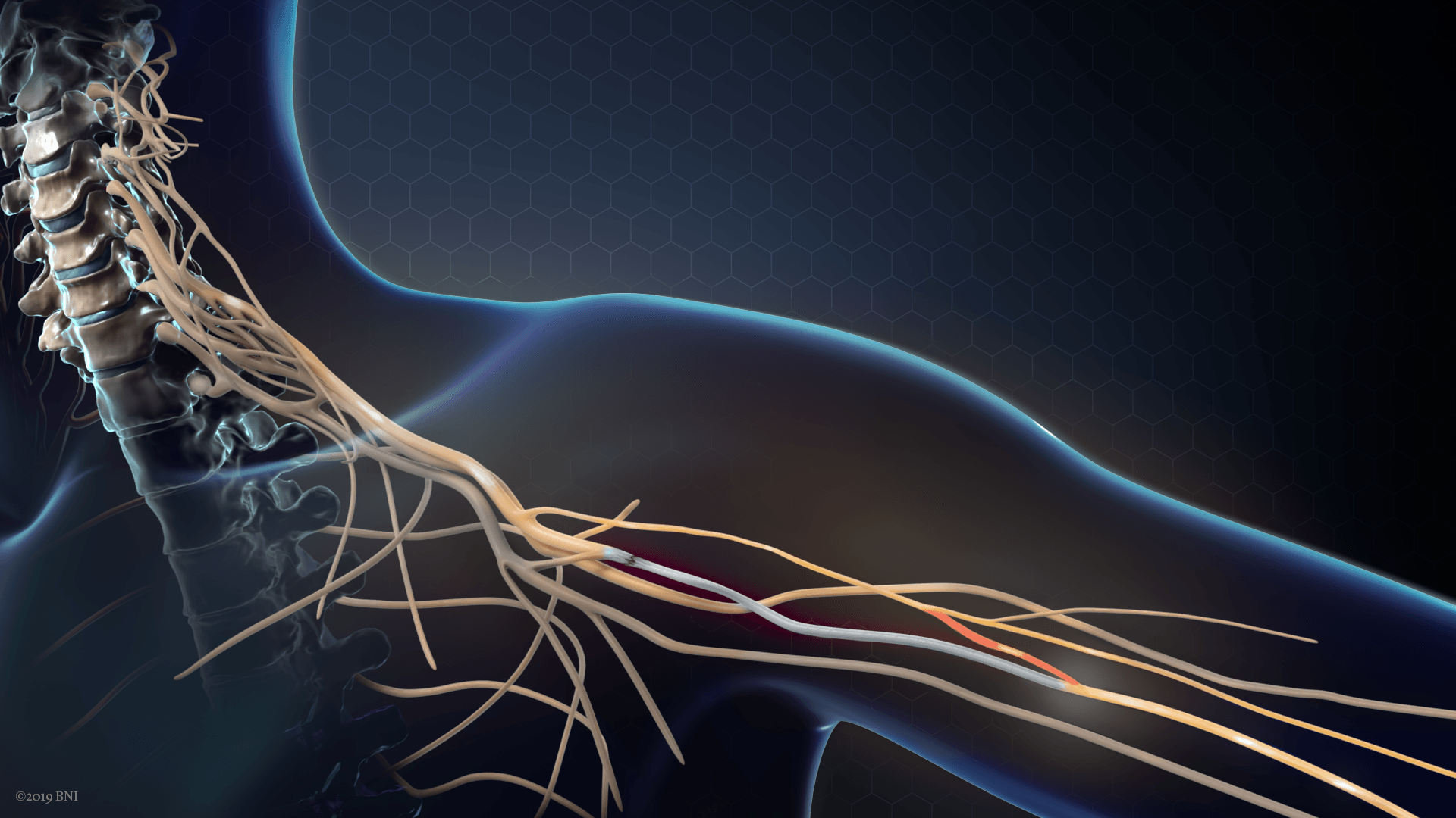
Peripheral nerves reside outside of the brain and spinal cord. They allow the central nervous system to communicate with the rest of the body. When damage occurs to peripheral nerves, people may experience chronic pain, muscle weakness, or paralysis.
Because body armor leaves legs and arms unprotected, peripheral nerve injuries represent a leading cause of disability among U.S. military veterans. However, they’re also common among civilians, as they can result from various accidents and health conditions.
Peripheral Nerve Surgery: The Challenges
Peripheral nerves can regrow after injury, and a neurosurgeon may be able to help promote this process through a nerve repair procedure. This either involves directly reconnecting the damaged ends of the nerve or inserting a piece of a nerve from elsewhere in the body, which is known as a nerve graft.
Although central nerves do not regrow, neurosurgeons are also beginning to harness the regenerative power of peripheral nerves to help restore some hand function and sensation in people with cervical spinal cord injuries. A surgeon can take a functioning nerve that branches off of the spinal cord above the level of the injury and transfer it to a nonfunctioning nerve below the injury. The functioning nerve can grow into the receiving nerve and assume its job.
“The problem is that someone might be weak or paralyzed, but it might only be from swelling,” explained Barrow neurosurgeon Rory Murphy, MD. “Swelling will generally get better over time, but an injury to the nerve itself is unlikely to get better on its own.”
Without being able to see the extent of the nerve damage, a surgeon faces a difficult decision of when and how to intervene.
When surgeons do perform a nerve repair or transfer, they have no objective way to monitor the patient’s recovery. Instead, the physicians depend largely on their clinical observations. Since peripheral nerves regenerate slowly—about an inch per month—it can take months or even years to learn that a nerve repair or transfer has failed. At that point, a revision surgery may no longer be an option.
“During that time, muscles start to atrophy,” Dr. Dortch said. “Once a muscle loses its input from a nerve, it starts to shrink—and that’s an irreversible process.”
Diffusion MRI: Monitoring Nerve Repair with Water Dynamics
As a solution, Dr. Dortch has proposed a type of magnetic resonance imaging called diffusion MRI. Not only is MRI less invasive than electromyography and nerve conduction studies, which measure the activity of muscles and nerves, but preliminary data suggests that it may offer earlier and more detailed information about nerve repairs.
Diffusion MRI specifically can reveal microscopic details about the structure of tissues in the body with help from a simple molecule: water.
As water molecules spread—or diffuse—through tissues, their movement is affected by interactions with the obstacles they encounter. In peripheral nerves, the obstacles are axons—the electrical wiring that facilitates communication between nerve cells.
“You can think of the axons in nerves as a bunch of stacked cylinders that are long,” Dr. Dortch said. “Water molecules will diffuse more freely along these cylinders than across them.”
This dependence based on direction is known as anisotropy. Diffusion that is uniform in all directions, on the other hand, is called isotropy.
The fewer axons that water molecules encounter, the more isotropic their diffusion becomes. As nerves regrow, water diffusion becomes more anisotropic. These changes in diffusion could serve as biomarkers, or objectively measurable biological characteristics, of nerve damage and regeneration.
DoD Grant: Collaboration at Barrow and Beyond
The DoD-funded project is a collaboration between Barrow and Vanderbilt University in Nashville, where Dr. Dortch began his work in peripheral nerve imaging. The grant will provide the institutions with $2 million over four years.

Vanderbilt researchers will focus on the preclinical studies, imaging nerve trauma and repair in rats over time. Dr. Dortch and Ping Wang, PhD, an associate professor in the Neuroimaging Innovation Center at Barrow, will perform translational studies in humans to move the imaging method toward clinical trial readiness.
The Barrow researchers will develop the clinical trial protocol, which will build upon Dr. Dortch’s work that was funded by a National Institutes of Health R01 grant. They aim to demonstrate consistency across different MRI scanners and evaluate whether diffusion MRI can identify failed nerve repairs earlier than existing methods in humans.
Validating the diffusion MRI method could be a game-changer for clinical trials of regenerative medicine. These trials would no longer depend on subjective clinical assessments, which are highly variable and slow to change.
Patients in this study will be scanned at both Barrow and Vanderbilt. Those at Barrow will undergo nerve repair procedures with Dr. Murphy.
Barrow offers this close integration between research and clinical care. Combining that with our access to unique patient populations and large clinical volumes makes this an ideal place for this type of clinical translation.
Richard Dortch, PhD
“We’ll scan them four times over a year and also follow them clinically,” Dr. Dortch said. “The hope is that the scans early after surgery are predictive of who ends up recovering and who does not.”
It’s a novel, and fitting, partnership: a neurosurgeon who specializes in repairing peripheral nerves and an engineer who has spent years developing a method to image them.
“Barrow offers this close integration between research and clinical care,” Dr. Dortch said. “Combining that with our access to unique patient populations and large clinical volumes makes this an ideal place for this type of clinical translation.”