Rathke’s Cleft Cyst
At a Glance
- A Rathke’s cleft cyst is a benign, fluid-filled pouch in the pituitary gland that develops during embryonic development.
- Most cysts are asymptomatic and discovered incidentally during MRI or CT scans.
- Symptomatic cases may cause visual disturbances, headaches, milky breast discharge, or diabetes insipidus.
- Treatment includes observation for mild cases or minimally invasive surgery to drain the cyst if symptoms are present.
Overview
A Rathke’s cleft cyst is a noncancerous, fluid-filled lesion that develops in the pituitary gland between the front and back lobes, known as the anterior and posterior lobes. The pituitary gland is a pea-sized organ at the base of the brain, producing essential hormones that regulate important bodily functions.
Rathke’s cleft cysts vary in size and appearance, and the fluid inside can be watery, gelatinous, or mucus-like. Many of these cysts are small and do not cause any symptoms. However, larger Rathke’s cleft cysts can press on nearby structures, like the pituitary gland or the optic chiasm, leading to headaches, hormonal imbalances, or visual disturbances.
What causes Rathke’s cleft cysts?
Rathke’s pouch is a structure that produces part of the pituitary gland during embryonic development. As development proceeds, the hollow center of the Rathke’s pouch shrinks into a cleft that recedes and disappears. However, if the pouch does not close completely and some of the tissue persists, a Rathke cleft cyst can form later in life.
The exact reason why remnants of Rathke’s pouch develop into a cyst in some people and not others isn’t fully understood. Still, we know that remnants from Rathke’s pouch cause Rathke’s cleft cysts and are present at birth.
Researchers have not linked Rathke’s cleft cysts to trauma, infection, or genetic inheritance. Instead, they’re considered an unusual byproduct of the formation of the pituitary gland.
Did you know?
Rathke’s pouch was first described by Martin Rathke, a German scientist from the 19th century, recognized as one of the founders of modern embryology.

Rathke’s Cleft Cysts Symptoms
Most Rathke’s cleft cysts are asymptomatic, meaning they don’t produce observable symptoms—in fact, doctors often discover them on brain imaging studies done for other reasons. However, if they grow large enough, these cysts can press on structures in the pituitary region and cause many problems.
Symptoms that may indicate you have a Rathke’s cleft cyst include:
- Headaches: As one of the most common symptoms, headaches often result when the cyst creates pressure within the sella turcica, a bony compartment at the base of the skull that houses the pituitary gland. Stretching of the outermost layer of the brain’s meninges, known as the dura mater, can also cause headaches.
- Endocrine dysfunction: Another symptom of a compressed pituitary gland due to a Rathke’s cleft cyst is hormonal imbalances, which can lead to hypopituitarism. This can feel like fatigue, weakness, low libido, or menstrual irregularities in women.
- Growth and puberty issues: This same disruption of pituitary hormones can also cause delayed puberty or growth abnormalities in children or adolescents.
- Visual disturbances: If a Rathke’s cleft cyst compresses your optic chiasm, which lives just above the pituitary gland, blurred vision or the loss of peripheral vision can occur.
- Hyperprolactinemia: If a Rathke’s cleft cyst compresses your pituitary stalk, it can disrupt dopamine delivery, leading to elevated prolactin levels. This, in turn, leads to hyperprolactinemia, which can cause milky discharge from the breasts called galactorrhea, or menstrual irregularities in women.
- Nausea and vomiting: If the cyst is large enough to raise pressure inside the skull, unexplained nausea and vomiting can occur.
When Rathke’s cleft cysts are symptomatic, the typical presentation of symptoms involves headaches, visual changes, and hormonal issues. These are known as the primary triad of Rathke’s cleft cyst symptoms.
Rathke’s Cleft Cysts Diagnosis
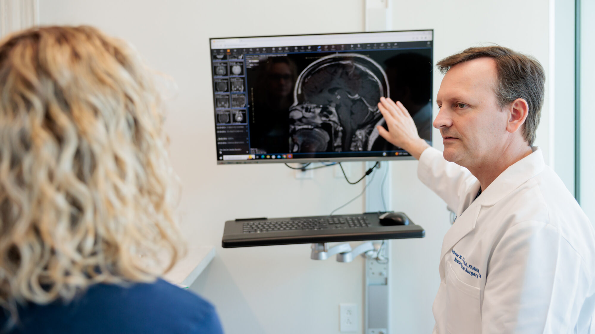
Physicians most often diagnose Rathke’s cleft cysts incidentally on brain imaging done for another reason. While magnetic resonance imaging (MRI) is the primary diagnostic tool, a complete evaluation and diagnosis often includes blood testing, vision testing, and, in some instances, surgical biopsy.
Doctors may use the following exams, tests, and imaging studies in the process of diagnosing a Rathke’s cleft cyst:
- Physical and neurological exam: First, your healthcare provider will ask about your symptoms, overall health, and family history. Next, they’ll complete an examination to assess neurological function, including your reflexes, coordination, strength, and sensation.
- Magnetic Resonance Imaging (MRI): Imaging studies are crucial in visualizing the brain and identifying abnormalities, and MRI is the gold standard for diagnosing Rathke’s cleft cysts. An MRI provides detailed images of the pituitary gland region. Rathke cleft cysts often appear as well-defined lesions within the sella turcica.
- Computed Tomography (CT): CT scans rely on X-rays to create detailed cross-sectional images of the brain when an MRI is not feasible or an additional evaluation is needed. While an MRI is superior to a CT scan for diagnosing a Rathke’s cleft cyst, a CT scan can still be helpful because it’s better at detecting traces of calcium. These traces can suggest a related but wholly different diagnosis: craniopharyngioma.
- Blood tests: A series of hormone panels done via a blood draw will check your hormone levels and look for imbalances in cortisol, prolactin, growth hormone, and thyroid hormones stemming from compression of your pituitary gland.
- Ophthalmologic exam: If your vision is affected due to pressure on the optic chiasm from a Rathke’s cleft cyst, an ophthalmologic exam will help assess its impact.
- Histopathology: In unclear cases where the Rathke’s cleft cyst is drained or removed, an examination after surgery can help confirm the diagnosis.

Rathke’s Cleft Cysts Treatment
The treatment of a Rathke’s cleft cyst depends on whether it is symptomatic or asymptomatic. You likely won’t need treatment if your Rathke’s cleft cyst was found incidentally and isn’t causing symptoms. When symptoms are noticeable, treatment often includes a surgical procedure.
Nonsurgical Treatments
Some Rathke’s cleft cysts don’t cause symptoms. If you’re not experiencing any symptoms, the best action may be to watch the cyst over time. This can look like regular monitoring via MRI scans to check for cyst growth or changes, and regular hormone panels to monitor pituitary functions.
Other nonsurgical approaches can include:
- Medical management of endocrine changes: If your Rathke’s cleft cyst causes hormone deficiencies, common treatments include hormone replacement therapy to manage your symptoms without directly treating the cyst itself. Examples include estrogen, progesterone, testosterone, or DHEA replacement, hydrocortisone for adrenal insufficiency, and levothyroxine for hypothyroidism.
- Headache management: If your cyst is small, headaches may still occur. Your care team will help you manage these with pain medication or migraine therapies.
Surgical Treatments
Approximately one in 10 people with Rathke’s cleft cysts need surgery to either drain the cyst or to remove as much of the cyst as possible. Rather than removing the entire cyst, which can damage the pituitary gland, a small portion of the membrane surrounding the cyst is removed, and its contents are drained. These contents are not infectious and will not harm your pituitary gland or the tissues around it.
- Transsphenoidal surgery: The most common surgical approach for a Rathke’s cleft tumor is transsphenoidal surgery. This is when a neurosurgeon reaches the cyst through the nasal passages and the sphenoid sinus using a small surgical instrument and an endoscope. Once your surgeon gains access to the cyst, they will create a hole in the sella turcica—the bone that cradles and protects your pituitary gland—so the cyst can be partially removed and drained. After removal, the surgeon will clean the cavity and seal it. Transsphenoidal surgery leaves no scar and minimizes the risk of complications.
- Cyst fenestration: A variation of transsphenoidal surgery, cyst fenestration involves a surgical opening that allows the cyst to drain continuously into the sphenoid sinus. Fenestration lowers the chance of fluid reaccumulating.
- Craniotomy: Although rare, your neurosurgeon may perform a craniotomy for larger or more complicated Rathke’s cleft cyst cases that are inaccessable via the transsphenoidal approach. During a craniotomy, a neurosurgeon will make an incision in the scalp, remove a portion of the skull, and access the pituitary gland to remove as much of the cyst as possible while using intraoperative imaging and highly specialized tools. This procedure is more invasive than transsphenoidal surgery and requires a longer recovery time.
Although surgery generally provides good outcomes, Rathke’s cleft cysts can recur. Long-term follow-up care with consistent MRIs and endocrine testing is critical even after successful surgery.
Common Questions
How common are Rathke’s cleft cysts?
Because most people don’t experience symptoms, it can be challenging to understand precisely how common Rathke’s cleft cysts are. They’re common as incidental findings, or when doctors find them during a procedure or test done for another reason. When found incidentally on MRIs, Rathke’s cleft cysts affect around 4% of American adults. Meanwhile, they’re found in approximately 3% of children, and are often smaller in size.
Only a small fraction of Rathke’s cleft cysts become large enough to cause problems, accounting for about 1% of all lesions diagnosed at the base of the brain.
Who gets Rathke’s cleft cysts?
While Rathke’s cleft cysts are not developmentally rare and they can occur at any age, symptomatic cases are most often detected in middle-aged adults—between the ages of 30 and 50—with a slightly higher incidence in women. The symptoms that most often present in this demographic are headaches, vision loss, and hormonal problems.
These cysts are much less common in children. Still, when they present, they can trigger delayed puberty, stunted growth, or visual symptoms.
What is the difference between a Rathke’s cleft cyst and a craniopharyngioma?
Rathke’s cleft cysts and craniopharyngiomas originate from remnants of Rathke’s pouch, but they’re fundamentally different in their behavior and impact.
A Rathke’s cleft cyst is noncancerous, mostly harmless, and often only found during incidental imaging studies. Meanwhile, a craniopharyngioma behaves more like a tumor, although it’s still benign. They are more aggressive than Rathke’s cleft cysts and require more extensive treatment because they can grow close to critical brain structures.
What is the prognosis for someone with a Rathke’s cleft cyst?
Overall, the outlook for someone with a Rathke’s cleft cyst is positive. Cysts without symptoms usually remain stable and don’t require treatment; just observation with periodic MRI scans. Cysts that have caused symptoms generally have a high rate of relief after surgery.
If the Rathke’s cleft cyst has caused pituitary hormone loss before surgery, you may continue to need lifelong hormone replacement therapy. Visual symptoms usually improve if treated early, but long-standing compression may lead to permanent visual deficits.
Unfortunately, Rathke’s cleft cysts have a significant rate of recurrence: studies suggest that around 30% of these cysts can reaccumulate fluid over time. However, not all recurrences will cause symptoms or require additional surgery.
Can Rathke’s cleft cysts be prevented?
It is not possible to prevent a Rathke’s cleft cyst from forming. The formation of a Rathke’s cleft cyst is not influenced by lifestyle, environment, or known risk factors. The only preventative measure is to monitor your Rathke’s cleft cyst once discovered to see if it remains stable or grows enough to require treatment.


