


Imaging Research Aims to Unravel How Parkinson’s Alters Brain Networks
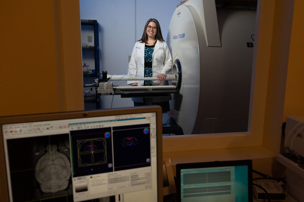
Ashley Stokes, PhD, a neuroimaging scientist at Barrow Neurological Institute, has received a $1.25 million grant from the National Institutes of Health to study Parkinson’s-related changes across different functional networks in the brain using an advanced MRI method.
The five-year R01 grant will support Dr. Stokes and her team as they work to accomplish two overarching goals. The first is to develop imaging-based biomarkers of Parkinson’s disease. Biomarkers are objective indicators that can show the presence of a disease, its progression, and how it’s responding to treatment. The second goal is to use these biomarkers to identify exactly how and where Parkinson’s wreaks havoc on the brain—especially beyond the dopamine circuit.
Parkinson’s is characterized by the gradual deterioration and death of nerve cells that produce dopamine. This chemical messenger plays a role in many functions, including muscle movements. Movement-related symptoms such as tremors, stiffness, and slowness are hallmark features of Parkinson’s.
However, less is known about how the disease affects non-dopaminergic neurons. Other neural circuits could hold the answers to why people with Parkinson’s also experience non-motor symptoms and medication side effects.
“For many patients, when they think about the symptoms that are bothering them the most, it’s not always the motor symptoms; it’s often a lot of the other symptoms,” Dr. Stokes said.
Cognitive changes, hallucinations, sleep and mood disorders, vision problems, and pain are just a handful of the non-motor symptoms that people with Parkinson’s may experience.
Unique Grant Funds New Research Direction
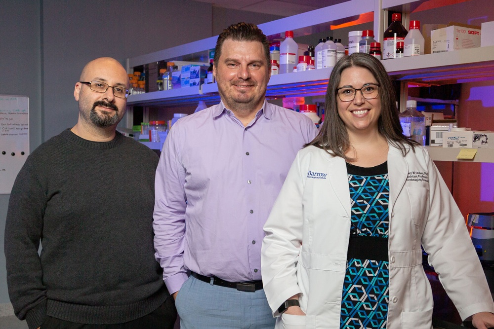
Non-motor symptoms and medication side effects in Parkinson’s disease are of particular interest to Fredric Manfredsson, PhD, an associate professor in the Department of Translational Neuroscience at Barrow. He has spent several years investigating the role of serotonin-producing neurons in levodopa-induced dyskinesias. These are uncontrollable muscle movements that develop in most patients taking the common Parkinson’s drug levodopa.
As part of this work, Dr. Manfredsson approached Dr. Stokes about collaborating to develop imaging-based biomarkers for Parkinson’s. Just days later, the National Institutes of Health announced a unique, new type of R01 grant. Named after a former NIH leader, the Stephen I. Katz Early-Stage Investigator Research Project Grant funds innovative ideas that represent new directions for early-stage investigators. The grant is unusual in that it doesn’t allow any preliminary data.
“Usually for an NIH grant, you need preliminary data just to apply,” Dr. Stokes said. “Your preliminary data shows that the project is feasible, that you are the person to do it, and that it’s worth doing.”
Because Drs. Stokes and Manfredsson had just started discussing the project, they hadn’t gathered data yet. Additionally, the project marks a change in research direction for Dr. Stokes, whose previous work has been in Alzheimer’s disease, brain tumors, and multiple sclerosis.
Overcoming Challenges to Develop Parkinson’s Biomarkers
While standard MRI shows abnormalities in brain tissues, functional MRI (fMRI) enables the measuring and mapping of brain activity. This method is not directly sensitive to neural activity, but it can detect subsequent changes in MRI signal through a mechanism called neurovascular coupling.
“It’s an indirect marker of brain activation,” Dr. Stokes said. “If I ask a participant to tap their finger, for example, then I can see where in the brain that lights up.”
But to reveal subtle changes in the brain related to Parkinson’s disease and evaluate emerging therapies for those changes, scientists need a direct biomarker. As a potential solution, Dr. Stokes proposed spin- and gradient-echo (SAGE)—an advanced multi-contrast fMRI method she has used in brain tumor research.
SAGE offers the advantage of providing information from both large and small blood vessel networks. These dual readouts will shape a more complete picture of how different parts of the brain communicate and how those signals are affected by Parkinson’s and its treatment. Maurizio Bergamino, PhD, an imaging scientist in the Stokes Laboratory, will help analyze that data.
“SAGE is, to my knowledge, unique to Barrow,” Dr. Stokes said. “I don’t know of any other sites that are using SAGE, especially not for fMRI.”
The team will apply SAGE to rat models of Parkinson’s, with Dr. Manfredsson guiding pharmacologic and chemogenetic experiments. These studies will involve modifying neuronal activity, both with gene therapy and drug therapy, and seeing what happens on imaging scans as a result.
“This work is essentially an extension of the programmatic focus of my group, aimed at understanding what changes in the Parkinson’s brain are responsible for the many non-motor symptoms affecting patients,” Dr. Manfredsson said. “In our work with Dr. Stokes, we are beginning to try to define the anatomical changes that happen in an animal model of Parkinson’s with the ultimate goal of translating these findings into human studies.”
Providing Hope for Patients with Parkinson’s
Although it is a preclinical grant, the researchers will compare the results of their preclinical studies to imaging scans of patients with Parkinson’s disease. For help with the clinical context, Dr. Stokes has teamed up with Barrow Parkinson’s neurologist Holly Shill, MD.

“The ability to find treatments that meaningfully slow or halt the decline of Parkinson’s disease has been hampered by our ability to accurately track its progression,” said Dr. Shill, who serves as director of the Muhammad Ali Parkinson Center at Barrow. “If successful, this study may lead to accurate methods to reliably track progression using simple imaging tools (MRI) available in most clinical settings.”
The opportunity to work closely with patients and positively impact their lives is what drew Dr. Stokes to the field of neuroimaging. It’s also why she brought her research to the Neuroimaging Innovation Center at Barrow several years ago.
“I hope that in the long term we could translate these methods into the clinical populations and give people answers for why they’re experiencing what they’re experiencing, whether it can be traced back to serotonin or another circuit that’s being impacted—and there could be treatment targets,” Dr. Stokes said. “I think that’s one of the unique things about Barrow—that we have the ability to forward translate and back translate. We have great resources on both sides.”