Advancing Precision
Barrow Neuroimaging Innovation Center
Advanced Technology
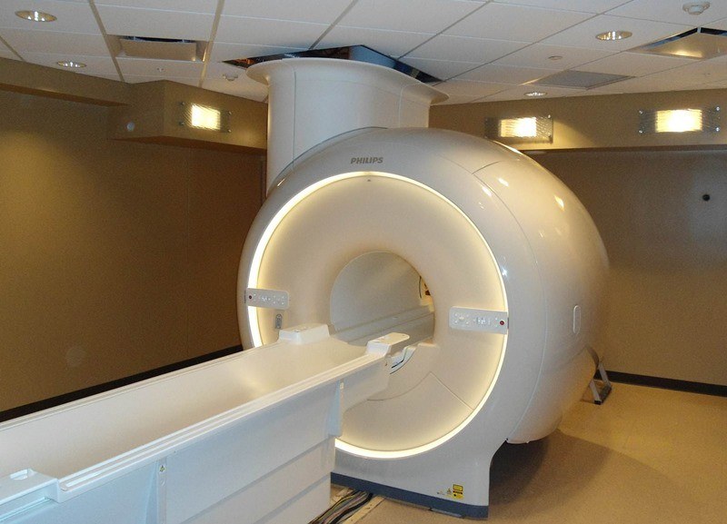
We operate a whole body 3-Tesla Philips Ingenia MRI scanner dedicated entirely to research. Our staff works with researchers from Barrow or any other institution wishing to utilize this scanner for their biomedical research.
The 3T MRI is equipped with a host of imaging capabilities to meet a variety of scanning needs, including:
- Anatomical Imaging. A variety of scans can be used to highlight almost any anatomy of interest. The latest technology provides high-resolution, accurate assessment of morphometry and volume.
- Functional MRI (fMRI). A variety of presentation hardware, including goggles and earphones, can be used to present stimulus to a subject in order to assess brain activation.
- Quantitative Flow and Diffusion Imaging. Advanced MR methods can measure and quantify motion as subtle as water diffusion as well as flow velocity in 3 dimensions, either in vivo or in physical models using a programmable MR compatible pump.
- Diffusion Tensor Imaging (DTI). In vivo measurement of the anisotropic diffusion of water is used to generate images of white matter tracts in the brain.
- Perfusion Imaging. The measurement of the effects of perfusion and contrast agents is a valuable tool for examining neuropathologies such as brain tumors and stroke. Endogenous contrast methods (e.g., arterial spin labeling, ASL) and contrast agent bolus tracking methods (dynamic susceptibility contrast and dynamic contrast enhanced MRI) are some of the methods available to researchers using the Center.
- Angiography. The visualization of vascular anatomy is important for the study of vascular disease, stenosis, and blood-flow occlusion. High-resolution in vivo imaging of vessels is possible with the use of specialized imaging techniques, contrast agents, or both.
- Susceptibility Weighted Imaging (SWI). SWI is a method that is sensitive to variations in a tissue’s local magnetic fields and can be used to enhance visualization of deoxyhemoglobin in veins, iron deposition in the brain, hemorrhages, microbleeds and calcification. SWI is currently used for a range of clinical applications including stroke, TBI and a range of neurodegenerative disorders.

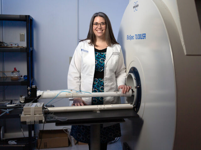
Our Laboratories
Our laboratories use advanced imaging hardware, cutting-edge methodology, and deep scientific insight to provide robust information for translating neuroimaging research into clinical impact.


Investigators


Sharmeen Maze RT(R)(MR)
Research MR Technologist
Sharmeen.Maze@CommonSpirit.org
(602) 406-2644
Cindy Moreno
Department Research Coordinator
Cindy.Moreno@CommonSpirit.org
(602) 406-4215
Gregory Turner, PhD
Preclinical Imaging Program Manager
Barrow Neuroimaging Innovation Center
350 West Thomas Road
Phoenix, Arizona 85013